Percutaneous coronary intervention (PCI), a cornerstone in the management of coronary artery disease, was first performed in 1977.1 Most procedural aspects have improved, producing better clinical outcomes; these aspects include superior stent design and implantation techniques, use of intracoronary (IC) physiology and imaging, and improved pharmacotherapy.2 However, the need for X-ray guidance, with its associated radiation exposure to patients and medical staff, remains. Interventional cardiologists have increased rates of cancers and cataracts compared to non-interventionalists.3 One case series documented brain tumours in interventional cardiologists and radiologists, with the majority being left-sided, closest to the X-ray emitter.4 Furthermore, heavy lead protection equipment worn to reduce radiation exposure can cause musculoskeletal injuries. Surveys of interventional cardiologists reported frequent orthopaedic injuries, with a statistically significant link between years worked, case volume, and incidence of musculoskeletal problems.35 Patients are at risk of stochastic (cancer) and deterministic (skin erythema and ulceration) effects. It has been estimated that the risk of a fatal malignancy is 5% per sievert of radiation exposure. In 2010, 0.59% of all cancers in the United Kingdom were attributed to diagnostic medical imaging radiation exposure.6
Robotic-assisted percutaneous coronary intervention (R-PCI) allows precise control of PCI equipment. It consists of a bedside drive, which includes a robotic arm attached to a motor drive and a single use cassette into which the assistant loads the PCI equipment (Figure 1A). The operator sits within a cockpit, which comprises three joysticks and a touchscreen, allowing for independent and precise control of the catheter, guidewire, and IC device (Figure 1B). The touchscreen has arrows allowing 1 mm movements backwards or forwards of all three devices, providing higher precision than can be achieved by the human hand. Manual inputs are required for arterial access, the diagnostic angiogram, and guide catheter engagement. The clinician may need to take over if the robot cannot complete the procedure. The two main R-PCI systems are R-One (Robocath) and CorPath GRX (Siemens Healthineers).
Initial experience with R-PCI has been encouraging. A meta-analysis of seven studies (2,230 patients) showed R-PCI had statistically significant reductions in air kerma (mean difference [MD] –442.3 mGy, 95% confidence interval [CI]: –675.9 to –208.8; p=0.0002), fluoroscopy time (MD –1.5 mins, 95% CI: –2.92 to 0.0; p=0.05), and contrast usage (MD –18.3 mL, 95% CI: –24.2 to –12.4; p<0.00001).7 Rates of major adverse cardiac events (MACE; odds ratio [OR] 0.9, 95% CI: 0.5-1.8), all-cause mortality (OR 1.0, 95% CI: 0.2-4.3), and myocardial infarction (OR 0.9, 95% CI: 0.4-1.9) were comparable between R-PCI and manual PCI (M-PCI).7 However, to our knowledge, no randomised clinical trials investigating R-PCI have been conducted, representing an evidence gap.
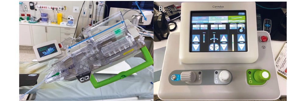
Figure 1. Robotic setup. Reproduced with permission from Dr J Leung.. A) Bedside console with robotic drive. B) Shielded operator cockpit with touchscreen and three joysticks to control the catheter, guidewire, and cardiac device.
Methods
Trial design and oversight
The Percutaneous coronary intervention using Assisted Robotic TechnologY (PARTY) trial was an unblinded, investigator-initiated, randomised controlled trial. The trial design and protocol have been published previously.8 Ethics approval (2022/ETH02267) and site-specific assessment authorisation (2022/STE03770) was granted by South Western Sydney Local Health District Human Research Ethics Committee, and the trial was prospectively registered with the Australian New Zealand Clinical Trials Registry (ACTRN12623000480684). All patients provided written informed consent. The trial was conducted in accordance with the Declaration of Helsinki. The first and last authors were responsible for drafting the manuscript and vouch for the accuracy of the trial database.
Patient selection
Patients aged 18-85 years who underwent invasive coronary angiography and PCI were eligible for recruitment. Elective outpatients and patients presenting with acute coronary syndrome were included. Recruitment necessitated patient allocation to a robot-capable catheterisation laboratory with a trained operator. Operator training included detailed lectures, simulator training, and five proctored cases. Patients were excluded if the operator deemed them or their coronary anatomy unsuitable for R-PCI, including chronic total occlusions, bifurcation lesions requiring an upfront two-stent strategy, or complex lesions requiring advanced calcium modification.
Randomisation and trial procedures
Study participants were randomised 1:1 to undergo R-PCI or M-PCI. R-PCI procedures were performed using the CorPath GRX robot. Randomisation was carried out using a web-based system after the diagnostic angiogram had been completed and the decision had been made to proceed to PCI. Prior to revascularisation, baseline patient demographics, medications, medical history, and pathology were collected.
Decisions related to the PCI procedure were at the operator’s discretion. Radial access was encouraged. Use of IC imaging was allowed, but as IC imaging catheters were not robot compatible, the robot was temporarily disengaged to allow a manual imaging run. The robot was then re-engaged to continue the PCI. IC imaging runs were recommended prior to lesion modification, after lesion modification, and after stenting. Operators were encouraged to use radiation settings and techniques to minimise radiation, including utilising a larger field size, minimising extreme angulation, and using filters/collimation to minimise radiation to non-essential regions. Fluoroscopy was recommended at 7.5 frames/second and cine acquisition at 10 frames/second. The assistant and scrub nurse were instructed to maximise their distance from the X-ray source. Use of ceiling-mounted radiation shielding and under-table lead protection was mandatory. The catheterisation table was raised as high as possible to maximise the patient-X-ray-source distance. Finally, source-image distance was minimised to reduce radiation scatter.
Trial endpoints
The aim of the PARTY trial was to investigate potential radiation benefits of R-PCI compared with M-PCI. The primary endpoint was patient radiation exposure, measured in microsieverts (μSv), recorded using a personal dosimeter placed at the patient’s left shoulder. Radiation exposure to the operator, assistant, and nurse were measured as secondary endpoints via dosimeters located in the chest pocket of the lead protection gown. The dosimeters used were the PM1610 X-ray and Gamma Radiation Personal Dosimeter (Polimaster). Clinical success was defined as <30% residual stenosis (by visual estimate) post-PCI and the absence of in-hospital MACE. R-PCI procedural success was defined as successful PCI completion without unplanned manual conversion. Manual conversion was categorised as either temporary manual assistance (intermittent manual inputs were required, but the robot still completed the procedure) or complete manual conversion. Temporary disengagement for IC imaging was not considered manual assistance. The total PCI time was measured from guiding catheter engagement to completion of the PCI procedure. Other procedural characteristics recorded were robotic setup time (defined as the time between opening the robotic equipment and the robot being ready for equipment loading), contrast usage, and radiation parameters (fluoroscopy time, digital stent enhancement software use, dose area product, and air kerma).
Safety outcomes, including MACE and other serious adverse events, were recorded. MACE was defined as cardiac death, clinically relevant myocardial infarction after coronary revascularisation (defined by the Fourth Universal Definition of Myocardial Infarction),9 or ischaemia-driven target vessel revascularisation. Bleeding complications were judged using the Bleeding Academic Research Consortium classification.10 Safety endpoints and adverse events were adjudicated by a blinded data safety and monitoring board (DSMB) of interventional and non-interventional cardiologists. Total equipment usage was recorded for future health economics analysis.
Operator workload was evaluated via the NASA Task Load Index (NASA-TLX) survey. This quantitative multidomain workload survey is the most cited survey-based workload measure.11 It has been utilised and validated in several fields, including medicine. The survey consists of six subdomains in which participants are asked to rate their physical, mental, and temporal demands, as well as their performance, effort, and frustration levels on a 21-point scale. An overall score (out of 100) is calculated. A higher score indicates a higher workload, except for performance, where a lower score indicates better performance.
Follow-up
At day three or hospital discharge (whichever occurred first), participants had their pathology recollected and interim clinical events recorded. Further clinical follow-ups occurred at days 30 and 365.
Statistical analysis
We initially calculated that a sample size of 300 patients would provide >99% power to detect an expected 20% reduction in patient radiation exposure, as was observed in a propensity score-matched analysis.12 Unfortunately, due to the early market withdrawal of the robot, the trial was terminated prematurely. Due to sufficient redundancy in the sample size calculations, a sample size of 150 patients still retained >90% power to detect the expected radiation reduction.
The trial data were analysed on an intention-to-treat basis. An as-treated analysis of the primary outcome was also performed. Continuous variables are presented as number of observations, with mean±standard deviation (SD) for normally distributed data or median and interquartile range (IQR) for non-normally distributed data. Categorical variables are summarised as the number and percentage frequencies. Continuous variables were analysed using an independent t-test or a non-parametric alternative (Mann-Whitney U test) depending on distribution assumptions. Categorical variables were compared using the chi-squared test. Two-sided 95% confidence intervals were calculated, and two-sided p-values<0.05 were considered statistically significant. Statistical analyses were performed using Stata statistical software, version 16 (StataCorp).
Results
Patient characteristics
Between June 2023 and January 2025, 452 patients undergoing coronary angiography were approached to assess their trial eligibility. Fifteen patients who required PCI, and consented, were deemed by the operator to be unsuitable for R-PCI (Supplementary Table 1). A total of 148 patients (163 lesions) were enrolled: 72 patients (78 lesions) were randomised to R-PCI and 76 patients (85 lesions) to M-PCI (Figure 2). Patient demographics and characteristics were equally balanced (Table 1). Most patients were male. Acute coronary syndrome was the most common clinical indication for PCI. The prescription of antiplatelets and cardiovascular risk factor-modifying treatments on discharge was high (Supplementary Table 2).
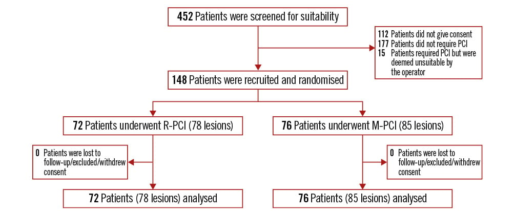
Figure 2. Trial CONSORT diagram. M-PCI: manual PCI; PCI: percutaneous coronary intervention; R-PCI: robotic-assisted PCI
Table 1. Baseline patient demographics
| Characteristic | R-PCI group n=72 | M-PCI group n=76 |
|---|---|---|
| Age, years | 64.0 (55.4-69.0) | 62.8 (56.0-69.9) |
| Male sex | 59 (81.9) | 63 (82.8) |
| Body mass index, kg/m2 | 28.1 (24.2-31.3) | 27.2 (25.1-31.3) |
| Hypertension | 46 (63.9) | 46 (60.5) |
| Hyperlipidaemia | 56 (77.8) | 56 (73.7) |
| Diabetes mellitus | 26 (36.1) | 26 (34.2) |
| Chronic renal failure* | 10 (13.9) | 8 (10.5) |
| Previous PCI | 24 (33.3) | 31 (40.8) |
| Previous MI | 18 (25.0) | 19 (25.0) |
| Previous CABG | 1 (1.4) | 5 (6.6) |
| Indication | ||
| Chronic coronary syndrome | 18 (25.0) | 15 (19.7) |
| Acute coronary syndrome | 54 (75.0) | 61 (80.3) |
| Current or previous smoker | 46 (63.9) | 47 (61.8) |
| Ejection fraction, % | 60 (45-60) | 60 (45-60) |
| Values are presented as n (%) or median (interquartile range). *Chronic renal failure was defined as an estimated glomerular filtration rate <60 mL/min/1.73 m2 for at least 3 months. CABG: coronary artery bypass grafting; MI: myocardial infarction; M-PCI: manual percutaneous coronary intervention; PCI: percutaneous coronary intervention; R-PCI: robotic-assisted percutaneous coronary intervention | ||
Procedures
Procedural characteristics are listed in Table 2. Most trial patients underwent single-vessel PCI for single-vessel disease via the radial approach. The median Synergy Between Percutaneous Coronary Intervention With Taxus and Cardiac Surgery (SYNTAX) scores were similar across treatment groups. Patients in the M-PCI group had a higher mean number and longer length of stents inserted, compared with those in the R-PCI group. IC imaging was utilised in 50% of total cases. However, more IC imaging was performed in the M-PCI group. The mean robotic setup time was 155.1±55.1 seconds.
Table 2. Procedural characteristics
| Characteristic | R-PCI group n=72 | M-PCI group n=76 | p-value |
|---|---|---|---|
| Number of vessels stented | NS | ||
| 1 vessel | 66 (91.6) | 68 (89.4) | |
| 2 vessels | 6 (8.3) | 6 (7.9) | |
| 3 vessels | 0 (0) | 2 (2.6) | |
| Vascular access | NS | ||
| Radial | 63 (87.5) | 70 (92.1) | |
| Femoral | 9 (12.5) | 6 (7.9) | |
| Vessels stented | 78 (100) | 85 (100) | |
| Left anterior descending artery | 37 (47.4) | 44 (51.7) | |
| Left circumflex artery | 15 (19.2) | 11 (12.9) | |
| Right coronary artery | 26 (33.3) | 29 (34) | |
| Bypass graft | 0 (0) | 1 (0) | |
| Pre-PCI stenosis, % | 80 (80-90) | 80 (80-90) | NS |
| SYNTAX score | 7 (5-12) | 8 (6-10) | NS |
| ACC/AHA lesion class | |||
| A | 2 (2.6) | 1 (1.1) | |
| B1 | 6 (7.7) | 4 (4.7) | |
| B2 or C | 70 (89.7) | 80 (94.1) | |
| Number of stents implanted | 1.3±0.7 | 1.8±0.9 | <0.01 |
| Stent length, mm | 25.0 (18-37.25) | 34.0 (23-56) | <0.01 |
| Intracoronary imaging | 23 (31.9) | 51 (67.1) | <0.01 |
| Intravascular ultrasound | 18 (78.3) | 37 (72.5) | NS |
| Optical coherence tomography | 5 (21.7) | 14 (27.5) | NS |
| Values are presented as n (%), mean±standard deviation, or median (interquartile range). N.B. 72 patients were randomised to the R-PCI group; however, a total of 78 lesions were treated. Similarly, 76 patients were randomised to the M-PCI group, with a total of 85 lesions treated. M-PCI: manual percutaneous coronary intervention; NS: not significant; PCI: percutaneous coronary intervention; R-PCI: robotic-assisted percutaneous coronary intervention | |||
Primary endpoint
Patient radiation exposure was reduced with R-PCI (Figure 3A), but this did not meet statistical significance in the intention-to-treat analysis (median R-PCI radiation 230.7 μSv [IQR 104-443.7] vs median M-PCI radiation 261.4 μSv [IQR 136.5-532.2]; p=0.14). However, statistical significance was met in the as-treated analysis (median 218.5 μSv [IQR 100-275.3] vs 264.7 μSv [IQR 136.3-534.8]; p=0.03).
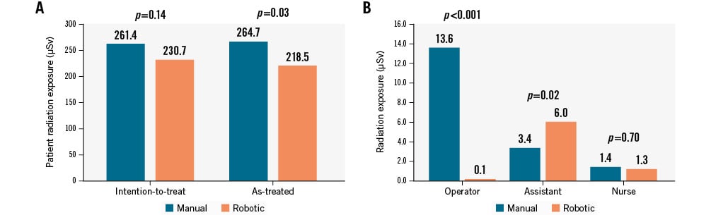
Figure 3. Radiation exposure. Radiation exposure to (A) patient (intention-to-treat and as-treated) and (B) operator, assistant, and nurse (intention-to-treat only). µSv: microsieverts
Secondary endpoints
PCI was successfully completed in all 148 patients (Table 3). Procedural success occurred in 61/72 of the R-PCI cases (84.7%) and in 67/78 of the R-PCI lesions (85.9%). Seven cases (9.7%) required manual conversion, while four cases (5.6%) required temporary manual assistance. The reasons for manual conversion included the following: inability to deliver equipment (n=3), irrecoverable guide disengagement (n=2), cassette malfunction (n=1), and coronary artery perforation (n=1). Most manual conversions occurred early in the recruitment period. Both R-PCI and M-PCI had comparable PCI times; however, use of contrast and digital stent enhancement were reduced with R-PCI (Table 3). Finally, whilst the total fluoroscopy time and dose area product were similar between treatment groups, R-PCI was associated with reduced air kerma (Table 3).
Within the R-PCI group, there was no difference between the total PCI time for cases performed with or without IC imaging. The median PCI time for R-PCI cases with IC imaging was 44 mins (IQR 33-69), and the median PCI time for R-PCI cases without IC imaging was 40 mins (IQR 30-56.5); p=0.21. However, IC imaging did result in an increased stent length implanted within the R-PCI group. The median stent length for R-PCI cases performed with IC imaging was 33 mm (IQR 25.5-54.5), whilst the median stent length for R-PCI cases performed without IC imaging was 23 mm (IQR 18-33); p=0.01.
Operator radiation exposure was reduced in the R-PCI group (Figure 3B), while radiation exposure to the assistant was increased with R-PCI; however, no difference in nurse radiation exposure occurred between groups (Figure 3B).
R-PCI operators experienced significant workload reduction across all domains of the NASA-TLX survey (Table 4). Importantly, there was no difference in frustration levels between groups. The median overall operator workload score was significantly reduced with R-PCI. The assistant experienced reduced mental, physical, and temporal demands during R-PCI procedures with reduced overall effort (Table 4). Interestingly, the nursing workload showed numerically higher effort levels, significantly increased frustration, and reduced performance with R-PCI.
Table 3. PCI outcomes
| Characteristic | R-PCI group n=72 | M-PCI group n=76 | p-value |
|---|---|---|---|
| Overall success | 72 (100) | 76 (100) | NS |
| Clinical success | 63 (87.5) | 64 (80.3) | NS |
| PPMI* | 12 (16.7) | 16 (21.1) | NS |
| Procedural success | 61 (84.7) | 76 (100) | |
| Manual conversion | 7 (9.7) | NA | |
| Manual assistance | 4 (5.6) | NA | |
| Serious procedural complications | 1 (1.4) | 0 (0) | NS |
| Total PCI time, mins | 40 (25.3-58.8) | 46 (31.0-55.8) | NS |
| Digital stent enhancement (number of runs performed) | 4 (2-6) | 7 (5-11) | <0.01 |
| Contrast use, mL | 75 (50-120) | 100 (70-130) | <0.01 |
| Radiation measures | |||
| Fluoroscopy time, mins | 14.7 (9.6-20.7) | 16.5 (10.3-23.5) | NS |
| Dose area product, dGy·cm2 | 209.4 (112.3-363.3) | 206.6 (139.8-394.9) | NS |
| Air kerma, mGy | 349 (222.3-699.3) | 504 (298.3-909.3) | <0.01 |
| Data are presented as n (%) or median (interquartile range). *As defined by the Fourth Universal Definition of Myocardial Infarction. MACE: major adverse cardiac events; NA: not applicable; NS: not significant; PCI: percutaneous coronary intervention; PPMI: periprocedural myocardial infarction | |||
Table 4. Comparison of NASA-TLX survey results between R-PCI and M-PCI
| Characteristic | R-PCI group n=72 | M-PCI group n=76 | p-value |
|---|---|---|---|
| Operator | |||
| Mental demand | 4.5 (3-6.75) | 6 (4-11) | 0.01 |
| Physical demand | 3 (2-5) | 7 (5-11) | <0.01 |
| Temporal demand | 4 (2-6) | 5 (3-9) | 0.03 |
| Performance | 3 (2-6) | 6 (4-7.75) | <0.01 |
| Effort | 4 (3-7.75) | 8.5 (6-11) | <0.01 |
| Frustration | 4 (2-9) | 6 (3-8) | 0.06 |
| Overall score | 12.5 (9.375-30) | 25 (11.25-45) | <0.01 |
| Assistant | |||
| Mental demand | 4 (2-6) | 7 (4-11) | <0.01 |
| Physical demand | 4 (1-6) | 7.5 (4-11) | <0.01 |
| Temporal demand | 3 (1-5) | 5 (3-9) | <0.01 |
| Performance | 4 (2-6) | 4 (3-6.75) | 0.59 |
| Effort | 5 (3-7) | 7 (3.25-11) | 0.02 |
| Frustration | 5 (3-11) | 5 (3-9) | 0.32 |
| Overall score | 15 (10-22.5) | 32.5 (15-50) | 0.06 |
| Nurse | |||
| Mental demand | 5 (2-10) | 4 (1-7) | 0.25 |
| Physical demand | 4 (2-8.75) | 4.5 (2-6.75) | 0.62 |
| Temporal demand | 3 (1-6) | 3 (2-6) | 0.38 |
| Performance | 4 (2-7.75) | 3 (1.25-6) | 0.03 |
| Effort | 8 (3-11) | 5 (2.25-10.75) | 0.06 |
| Frustration | 3 (1-6) | 2 (1-4) | 0.02 |
| Overall score | 15 (5-32.5) | 13.75 (4.375-25) | 0.16 |
| Data are presented as median (interquartile range). M-PCI: manual percutaneous coronary intervention; NASA-TLX: NASA Task Load Index; R-PCI: robotic-assisted percutaneous coronary intervention |
|||
Safety endpoints
Serious procedural complications occurred in only one R-PCI patient (n=1, 1.4%) (Table 3). This was a coronary perforation during post-dilatation, which required covered stent insertion and was deemed not robot-related by the DSMB. Periprocedural myocardial infarction (PPMI) occurred in 12/72 (16.7%) of the R-PCI cases and 16/76 (21.1%) of the M-PCI cases. No cardiac deaths or deaths from any cause occurred in either group during hospital admission or at 30-day follow-up.
Discussion
This was the first randomised controlled clinical trial investigating R-PCI. We showed that patient radiation exposure was numerically reduced with R-PCI. This reduction became statistically significant in the as-treated analysis. The R-PCI operators experienced a 99.3% reduction in radiation exposure. This reduction is greater than the 95.2% reduction seen in the first large-scale, non-randomised study of R-PCI performed by Weisz et al in 2013.13 R-PCI was associated with other procedural benefits, including reduced contrast usage, less frequent use of digital stent enhancement software, and reduced air kerma (Central illustration). These procedural benefits are likely due to improved visualisation provided to the operator by sitting within the cockpit closer to the fluoroscopy screen. As a result, fewer contrast injections, less fluoroscopy time, and fewer digital stent enhancement runs were required to confirm IC device location. The procedural table can be raised higher than in a manual case, and a larger field size can be used, also reducing radiation.
Interestingly, the assistants in R-PCI cases were exposed to 76% more radiation than in M-PCI cases. This is perhaps because, during R-PCI, the assistant stands closer to the X-ray source and is not shielded by the operator as they would be in an M-PCI case. However, the median radiation exposure of the R-PCI assistants was lower than the median radiation exposure of the M-PCI operators (3.4 μSv vs 13.6 μSv). There was no difference in the radiation exposure of nurses between R-PCI and M-PCI cases. This is likely because nurses stand farthest away from the X-ray source; thus, any potential radiation difference with R-PCI is minimised.
Operator workload was significantly reduced in our study. With R-PCI, the operator sits within the shielded cockpit, thus avoiding prolonged periods of standing at the catheterisation table wearing lead protection equipment. This is reflected in the overall reduced workload experienced by the operator, particularly in the physical, mental, and temporal domains. The precision provided by R-PCI may have explained the operators’ perceptions of improved performance and reduced effort. The assistants also experienced reduced mental, physical, and temporal demands; however, the nurses experienced increased effort and frustration with reduced performance. This may be related to the additional nursing responsibilities during an R-PCI case. Finally, despite the extra time required to set up and engage the robot, there was no statistically significant difference in the total PCI time.
Serious procedural complications were rare. The coronary perforation in the R-PCI group was related to post-dilatation with a non-compliant balloon, rather than a robot-related complication. Rates of PPMI were similar between the R-PCI and M-PCI groups (Table 3). Our observed PPMI rate of 18.9% was comparable to previous reports.14 Using the Society for Cardiovascular Angiography and Interventions definition,15 our observed PPMI rate was even lower (3.4%). Adjudicating PPMI in patients whose preprocedural biomarkers are already elevated, or rising, is challenging. This was highlighted in our study given the high proportion of acute coronary syndrome patients recruited (n=110, 74.3%). This, coupled with the high proportion of complex lesions intervened on in the trial (92% ACC/AHA lesion classification B2 or C), could contribute to PPMI. Importantly, other MACE endpoints such as all-cause death, cardiac death, stent thrombosis, or target lesion revascularisation did not occur.
Our results are comparable to, or better than, previously published data. Compared with the meta-analysis of non-randomised data performed by Gupta et al, our observed R-PCI time, contrast use, and air kerma were similar or better, and our mean differences between R-PCI and M-PCI characteristics were greater.7 Our mean R-PCI time was 45 mins (mean between-group difference –4.0 mins) compared with 53.7 mins in the meta-analysis (mean difference 2.37 mins). The mean contrast use in our R-PCI cohort was 87.6 mL (mean between-group difference –25.8 mL) compared with 137.2 mL (mean difference –18.3 mL) reported in the meta-analysis. Finally, the mean air kerma in our R-PCI group was 488.5 mGy (mean between-group difference –233.5 mGy), whereas the meta-analysis reported a mean air kerma of 1,275.2 mGy (mean difference –442.7 mGy). The large difference in results, particularly the air kerma, could be because of the three studies included in the pooled analysis, one was performed in 2014, and one investigated robot-assisted chronic total occlusion (CTO) PCI. Older X-ray systems may not have the technological advancements that newer systems utilise to minimise radiation exposure. Furthermore, CTO PCI procedures are more complex, leading to longer procedure times and more radiation exposure.
Our procedural success rate for R-PCI was 84.7% with 9.7% (n=7) manual conversion and 5.6% (n=4) temporary manual assistance. Jaffar-Karballai and colleagues reported a median technical success rate of 93.8% (IQR 89.1-99.1) and a median partial manual assistance rate of 0% (IQR 0-7.6).16 Their definition of technical success allowed for partial manual assistance without compromising a “successful” robotic procedure. Using this definition, our technical success rate was 90.3%. Procedural success was high despite relative operator inexperience, requiring only five proctored cases before being eligible for trial participation.
The major strength of our trial is that it is the first randomised controlled trial investigating R-PCI. Consequently, potential confounders such as lesion complexity were equally balanced between treatment groups. Our trial was the first to record personalised radiation exposure for the whole medical team as well as the patient. Another first in R-PCI studies is our investigation and quantification of potential workload benefits for the medical team. The broad inclusion criteria facilitated recruitment of a diverse cohort, encompassing various clinical indications and complexities. We have completed clinical follow-up to 30 days, and longer-term follow-up is ongoing.
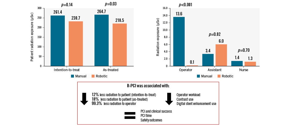
Central illustration. First randomised clinical trial investigating robotic-assisted percutaneous coronary intervention. µSv: microsieverts; PCI: percutaneous coronary intervention; R-PCI: robotic-assisted PCI
Limitations
There were several limitations to our trial. First, the unexpected early withdrawal of the robotic system reduced the trial recruitment period and, hence, the final sample size. This, together with a smaller than expected radiation difference, caused the trial to be underpowered and may have contributed to the non-significant primary endpoint. The lack of blinding may have introduced bias. The trial was performed at a single centre, which may also limit generalisability of results. The relative operator inexperience could have potentially diluted the radiation-saving benefits of R-PCI and increased rates of manual assistance or conversion. Whilst we endeavoured to recruit patients with varied clinical and procedural complexities, the lesions included were relatively simple. Most patients received single-vessel PCI and had a low SYNTAX score. Whilst safety endpoints and complications were independently assessed by a blinded DSMB, we did not have a core lab available to analyse angiogram images or other procedural data. Furthermore, whilst the NASA-TLX survey is a quantitative workload assessment, it is still subjective and prone to recall bias as it is completed post-procedurally. The current cost of the robot and associated consumables may be prohibitive to many centres. We hope to complete a subsequent health economics analysis to assess the cost-effectiveness of R-PCI. Finally, the current generation of cardiac robots is not compatible with certain PCI equipment, e.g., IC imaging catheters and over-the-wire devices; they are also unsuitable for lesions requiring multiple simultaneous balloon or stent deployments. This limits the wider uptake of R-PCI until these shortcomings are addressed.
The significant difference in IC imaging use between R-PCI and M-PCI is notable. It is well established that IC imaging use is associated with reduced procedural complications.17 The procedural and longer-term clinical benefits of IC imaging are more evident, with both the 2024 European Society of Cardiology Guidelines for the Management of Chronic Coronary Syndromes18 and the 2025 ACC/AHA Guideline for the Management of Patients with Acute Coronary Syndrome19 endorsing IC imaging as a Class I, Level of Evidence A recommendation to guide complex PCI. However, both guidelines were published after trial commencement.
IC imaging use may increase procedural time and, hence, radiation and contrast use, particularly with optical coherence tomography (OCT).20 This may explain the differences in PCI time, radiation, and contrast use identified in our study. However, most IC imaging cases utilised intravascular ultrasound (IVUS) (Table 2), which requires minimal additional radiation and contrast. Of the imaging cases in each group, there was no statistical difference between the proportion of cases performed with OCT or IVUS (Table 2). Furthermore, within the R-PCI group, using IC imaging did not lead to a statistically significant increase in procedure time compared with cases where IC imaging was not used. The rate of IC imaging use in the R-PCI group (31.9%) is higher than that reported in current practice in the United States (10.5%)21 or Australia (2.2%).22 The higher usage of IC imaging in the M-PCI arm could explain the greater number and longer length of stents implanted in that group. This is supported by the fact that the median stent length in the R-PCI group was statistically significantly longer when IC imaging was used. However, R-PCI can provide more accurate lesion and stent length assessment, which can potentially avoid unnecessary extra stents being deployed.23 This could also contribute to the smaller number and shorter length of stents implanted in the R-PCI group.
One potential application for R-PCI is a “hybrid” PCI case, e.g., during CTO PCI. These procedures were excluded from our study. Hirai and colleagues performed 49 robot-assisted hybrid single-vessel CTO PCI, where the CTO was crossed manually but the rest of the PCI procedure was completed robotically. They achieved similar success rates in the robot-assisted CTO PCI group compared with the manual control group, with comparable safety.24 Operators in the robot-assisted CTO PCI group spent 48% of the total procedure time within the shielded cockpit, suggesting that significant radiation and orthopaedic workload reductions could be achieved with robot-assisted CTO PCI. A hybrid approach may allow the benefits of R-PCI (namely reduced radiation and physical strain) to be experienced whilst not compromising best contemporary practices for PCI (utilising IC imaging or other devices incompatible with the robot).
Conclusions
In the first randomised clinical trial investigating R-PCI, we demonstrated reduced patient radiation exposure, which met statistical significance in the as-treated population. Contrast use, air kerma, and use of stent enhancement software were reduced with R-PCI. Operator radiation exposure and their perceived workload were significantly reduced with R-PCI. Importantly, safety and efficacy outcomes were comparable between R-PCI and M-PCI. However, there are key limitations that must be addressed before this technology can be more widely utilised.
Impact on daily practice
Robotic-assisted percutaneous coronary intervention (R-PCI) is an evolving field of interventional cardiology. In this first randomised clinical trial of R-PCI, we have demonstrated radiation and procedural benefits with comparable safety outcomes versus manual percutaneous coronary intervention. However, there are key limitations that must be addressed.
Acknowledgements
We would like to thank all patients who participated in the trial. We thank Associate Professor Melissa Leung and Dr Tuan Nguyen for their assistance with study analysis. A special thank you to all the staff in the Liverpool Hospital cardiac catheterisation laboratory for their work, as well as the indispensable efforts from Siemens Healthineers (particularly Scott Sawyer and Jake Duong) for their training and technical support during the trial.
Funding
The cost of the CorPath GRX robot and associated consumables was supported by philanthropic donation via the Ingham Institute for Applied Medical Research. We also acknowledge the support and funding from the Ingham Institute for Applied Medical Research, University of New South Wales and the National Heart Foundation, without which this research would not have been possible. J. Leung is supported as a PhD candidate through the University of New South Wales and also by a National Heart Foundation PhD Scholarship (ID 107684).
Conflict of interest statement
The authors have no relevant conflicts of interest to declare.