Skip to content
The reference academic journal by and for the Asia-Pacific interventional cardiology community
AsiaIntervention

AsiaIntervention

  • Current issue
  • Archives
  • How to submit
    • Authors guidelines
    • Submit your paper
    • Reviewers guidelines
  • Services
    • Advertising
    • Article reprints
    • Publication calendar
    • Rights & Permissions
  • About the journal
    • Overview
    • Editorial Board
    • Masthead
  • Contact us
Volume 6 – Number 1 – July 2020

Successful management of shortening of overlapping segment and subsequent restenosis at the gap between two second-generation stents in the left anterior descending artery

AsiaIntervention 2020;6:56-57 | 10.4244/AIJ-D-18-00060

Nozomi Kotoku1, MD, PhD; Yuki Ishibashi1, MD, PhD; Yasuhiro Tanabe1, MD, PhD; Akiyuki Kotoku2, MD, PhD; Hiroshi Yamazaki1, MD; Shingo Kuwata1, MD, PhD; Takanobu Mitarai1, MD; Kazuaki Okuyama1, MD, PhD; Kihei Yoneyama1, MD, PhD; Takumi Higuma1, MD, PhD; Tomoo Harada1, MD, PhD; Yoshihiro J. Akashi1, MD, PhD

1. Division of Cardiology, Department of Internal Medicine. St. Marianna University School of Medicine, Kawasaki, Kanagawa, Japan; 2. Division of Radiology, St. Marianna University School of Medicine, Kawasaki, Kanagawa, Japan

A 74-year-old man with silent myocardial ischaemia underwent percutaneous coronary intervention to overlap a 3.0×38 mm everolimus-eluting stent (EES; XIENCE Alpine™, Abbott Vascular, Santa Clara, CA, USA) and a 2.5×16 mm everolimus-eluting platinum chromium stent (PtCr-EES; Promus PREMIER™, Boston Scientific Corp., Marlborough, MA, USA) in the mid-portion of the left anterior descending artery (LAD) (Moving image 1). Nine months after the stent implantation, the patient was readmitted to our hospital due to exertional chest pain. Coronary angiography (CAG) revealed significant restenosis at the mid-portion of the tortuous LAD segment (Moving image 2). Coronary computed tomography showed a distal stent fracture and shortening of the overlapping segment (Moving image 3). Optical coherence tomography (OCT) was also performed for the detailed examination. No significant luminal narrowing of the fractured segment was found (OCT, 3.97 mm2); whereas the minimum lumen area of the gaps between the two stented segments was 1.07 mm2 (OCT) (Moving image 4). A 3.0×12 mm PtCr-EES was successfully overlapped at the gap segment, from the proximal to the previously overlapped segment (Moving image 5). Follow-up CAG at 9 months after the second procedure resulted in no clinical events.

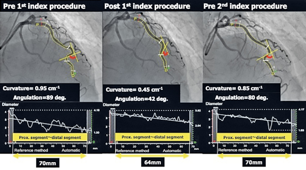
In the present case, the quantitative CAG analysis suggested that shortening of the overlapping segment and the gap between the two second-generation DESs might have occurred due to 1) straightening of the vessel curves after metallic stent implantation and 2) geometric changes caused by stent placement (Figure).

After the gap, caused by continuous stress during cardiac cycle, was created between the two second-generation DESs in the LAD, mechanical stress imposed on the stent edges would cause vessel wall injury and inflammation, characterised by the deposition of platelets and fibrin, as well as adhesions of circulating neutrophils and macrophages, which might lead to restenosis.

Previous studies reported that the PtCr-EESs with higher bending flexibility had greater fracture resistance against more severe angulation ranges than the other second-generation drug-eluting stents. In this case, the PtCr-EES was thus selected for significant restenosis at the gap between the two metal stents.

Conflict of interest statement

The authors have no conflicts of interest to declare.

Share

Supplementary data

https://www.asiaintervention.org/wp-content/uploads/2021/07/AIJv6i1_Kotoku_video1.mp4

Moving image 1. CAG after percutaneous coronary intervention and stenting of the LAD.

https://www.asiaintervention.org/wp-content/uploads/2021/07/AIJv6i1_Kotoku_video2.mp4

Moving image 2. CAG showing significant restenosis at the mid-portion of the tortuous LAD segment.

https://www.asiaintervention.org/wp-content/uploads/2021/07/AIJv6i1_Kotoku_video3.mp4

Moving image 3. Coronary computed tomography showing the distal stent fracture and shortening of overlapping segment.

https://www.asiaintervention.org/wp-content/uploads/2021/07/AIJv6i1_Kotoku_video4.mp4

Moving image 4. OCT showing the distal stent fracture and the gaps between the two stented segments.

https://www.asiaintervention.org/wp-content/uploads/2021/07/AIJv6i1_Kotoku_video5.mp4

Moving image 5. CAG after a PtCr-EES successfully overlapped the gap segment.

Volume 6 - Number 1

View full issue

Download this article
Authors
  • Akiyuki Kotoku
  • Hiroshi Yamazaki
  • Kazuaki Okuyama
  • Kihei Yoneyama
  • Nozomi Kotoku
  • Shingo Kuwata
  • Takanobu Mitarai
  • Takumi Higuma
  • Tomoo Harada
  • Yasuhiro Tanabe
  • Yoshihiro J. Akashi
  • Yuki Ishibashi
AsiaIntervention
  • Readers
    • Archives
    • Subscribe to the newsletter
    • Contact us
  • About the journal
    • Overview
    • Editorial Board
    • Masthead
  • Services
    • Advertising in AsiaIntervention
    • Article reprints
    • Publication calendar
    • Rights & Permissions
  • Authors
    • Authors guidelines
    • Submit your paper
  • Legal
    • Disclaimer
    • Cookies Policy
    • Privacy Policy
    • Legal Notice
  • Follow us
    • Facebook
    • X
    • LinkedIn
Online ISSN 2491-0929 - Print ISSN 2426-3958
© 2015-2025 Europa Group - All rights reserved