A 67-year-old female presented with ST-segment elevation myocardial infarction due to a heavily calcified lesion in the mid-right coronary artery (RCA) (Figure 1A). The initial intervention was performed via a 6 Fr femoral access.
During predilatation, a 2.5×8 mm non-compliant (NC) balloon failed to track through the lesion (Figure 1B). Subsequent attempts to withdraw the balloon resulted in shaft fracture and simultaneous loss of the guidewire. This critical complication left the balloon fragment entrapped within the calcified stenosis (Figure 1C), compromising coronary flow, resulting in Thrombolysis in Myocardial Infarction (TIMI) 1 flow.
After upgrading the vascular access site to an 8 Fr sheath and a 7 Fr Judkins right 3.5 guiding catheter, a second “buddy” guidewire was carefully navigated past the entrapped balloon. An initial retrieval attempt using a 2 mm Amplatz Goose Neck coronary snare (Medtronic) failed owing to the impacted position of the device and poor trackability (Figure 1D). To restore perfusion and create a working channel, intracoronary nitroglycerine was administered, and a 1.5×8 mm NC balloon was inflated alongside the fragment, successfully re-establishing TIMI 3 flow.
A 2.0×8 mm NC “buddy” balloon was then advanced over the wire parallel to the entrapped balloon and inflated at low pressure (4-6 atm) to create a significant frictional force against the entrapped device, effectively coupling the two balloons together. A gentle “push-and-pull” technique was applied using the inflated buddy balloon to gain traction and dislodge the fractured segment. This controlled traction, transmitted via the inflated buddy balloon, served to dislodge the impacted segment from the calcified stenosis. This manoeuvre successfully allowed the controlled withdrawal of the fractured balloon into the more spacious proximal RCA (Figure 1E). From this favourable position, the balloon was easily captured with a 4 mm Amplatz Goose Neck snare and removed (Figure 1F–Figure 1I).
The underlying calcified lesion was predilated and treated with two overlapping drug-eluting stents (3.0×48 mm and 3.5×20 mm), achieving an excellent final angiographic result with TIMI 3 flow (Figure 1H). The entire procedure is demonstrated in Moving image 1.
The management of a fractured and entrapped coronary balloon, particularly with the loss of guidewire access, presents a significant challenge. As outlined in established algorithms, when the fragment is located within the coronary artery but outside the guiding catheter, snaring is the recommended first-line approach12. In our case, this was initially unsuccessful because of balloon impaction within the calcified lesion. Preventing such complications is paramount and includes meticulous lesion preparation; in heavily calcified lesions, this ideally involves intravascular imaging and consideration for atherectomy. Furthermore, initiating predilatation with a smaller, highly trackable balloon could mitigate the risk of device entrapment. Instead of pursuing less favourable strategies, such as stenting over the fragment or emergency surgery, we employed this novel buddy balloon technique. This manoeuvre successfully created the necessary space and leverage to reposition the fragment, facilitating its eventual retrieval with a conventional snare.
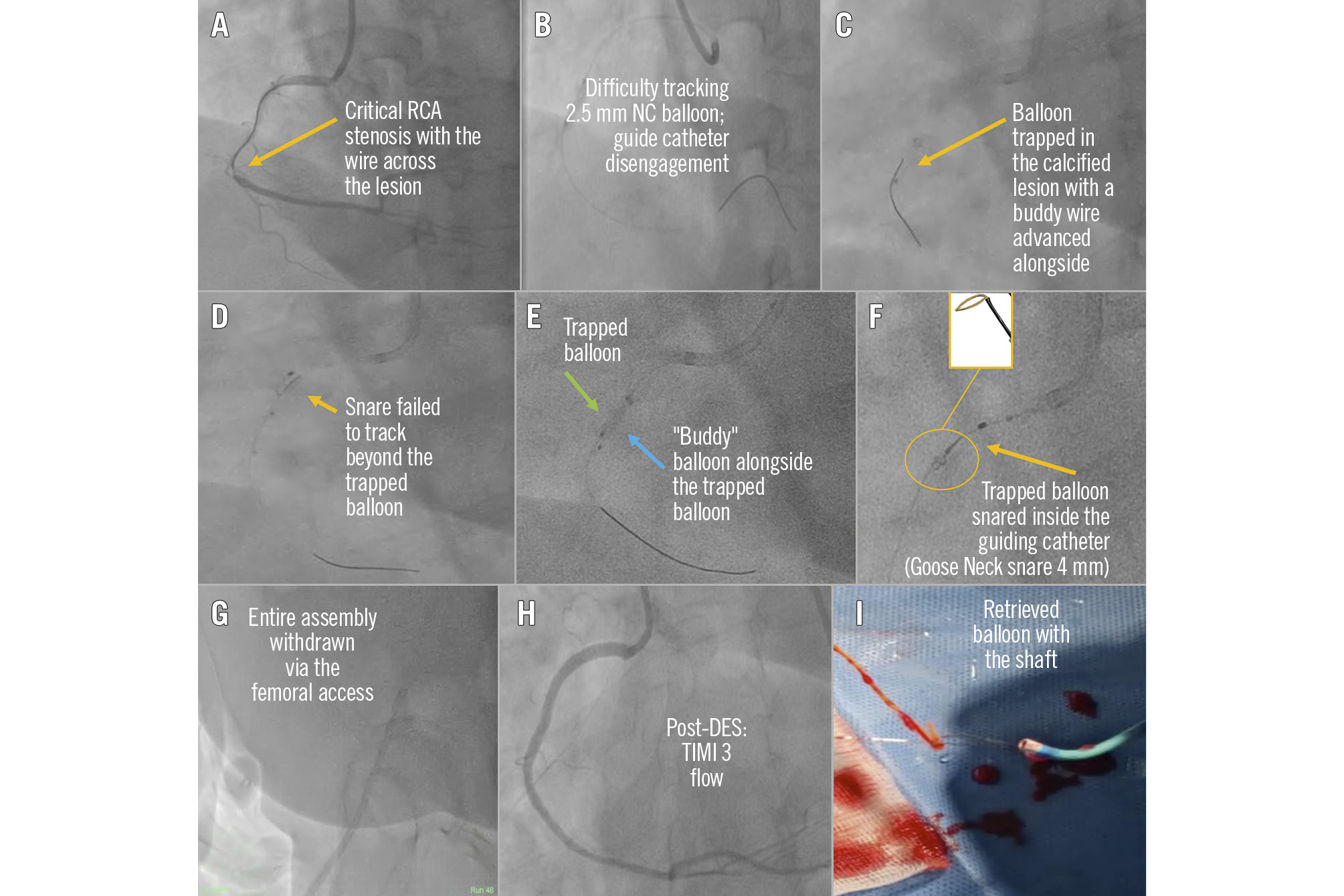
Figure 1. Percutaneous retrieval of a wireless, fractured coronary balloon. A) Baseline critical right coronary artery (RCA) stenosis with a wire across the lesion. B) Difficult balloon tracking, leading to guide catheter disengagement. C) The fractured balloon shaft is entrapped in the lesion, with a “buddy” guidewire advanced alongside it. D) Failed initial snaring attempt. E) “Buddy” balloon inflated parallel to the trapped balloon. F) Repositioned balloon snared in proximal RCA (inset showing 4 mm Amplatz Goose Neck snare). G) Entire assembly withdrawn via the femoral access. H) Excellent final angiographic result after stenting. I) Retrieved hardware. DES: drug-eluting stent; NC: non-compliant; TIMI: Thrombolysis in Myocardial Infarction
Consent statement
The authors confirm that informed consent was obtained from the patient for the intervention described in the manuscript and for the publication of their data, including all images.
Conflict of interest statement
The authors have no conflicts of interest to declare regarding the submitted work.