Skip to content
The reference academic journal by and for the Asia-Pacific interventional cardiology community
AsiaIntervention

AsiaIntervention

  • Current Issue
  • Archives
  • How to submit
    • Authors guidelines
    • Submit your paper
  • Services
    • Advertising
    • Article reprints
    • Publication calendar
    • Rights & Permissions
  • About the journal
    • Overview
    • Editorial Board
    • Masthead
  • Contact us
Volume 9 – Number 1

Intimal flap sealing and healing with perfusion balloon in spontaneous coronary artery dissection

AsiaIntervention 2023;9:64-65 | 10.4244/AIJ-D-22-00054

Yusuke Oba1, MD, PhD; Hiroshi Funayama1, MD, PhD; Keisuke Narita1, MD, PhD; Hajime Shinohara1, MD; Kazuomi Kario1, MD, PhD

1. Department of Cardiovascular Medicine, School of Medicine, Jichi Medical University, Shimotsuke-shi, Japan

A 70-year-old woman with no history of cardiovascular disease was admitted to our hospital for inferior ST-segment elevation myocardial infarction. Urgent coronary angiography (CAG) revealed severe stenosis with Thrombolysis and Myocardial Infarction (TIMI) flow grade 1 in the right coronary artery (RCA) (Figure 1A, Moving image 1). Due to anterograde flow impairment and ongoing ischaemia, we dilated the lesion with a 2.0 mm balloon catheter (Sapphire II; OrbusNeich). Intravascular ultrasound (IVUS) imaging (AltaView; Terumo) clearly showed an intimal flap (Figure 1B, Moving image 2) and a massive intramural haematoma (IMH) at the proximal end of the flap. We diagnosed the patient with retrograde spontaneous coronary artery dissection (SCAD). We performed a long inflation, for 5 min, with a 2.5 mm perfusion balloon (PB) (Ryusei; Kaneka Medix) (Figure 1C). Satisfactory acute gain was achieved (Figure 1D, Moving image 3) and TIMI flow grade 3 was obtained without IMH propagation. On the 14th day of hospitalisation, CAG revealed moderate stenosis in the RCA (Figure 1E), even though the level of fractional flow reserve (FFR) was 0.95. Optical coherence tomography (OCT) and IVUS clearly showed the healed flap without IMH propagation (Figure 1F, Figure 1G, Moving image 4, Moving image 5). Therefore, we decided to defer from performing additional percutaneous coronary interventions. Three months later, the patient’s coronary computed tomography showed the lesion to be completely healed (Figure 1H, Figure 1I). Since a PB has the advantage of maintaining blood flow during balloon inflation, repeated dilatation of the target lesion for longer than several minutes was possible. Although PB have been used as bailout devices in cases of vessel perforation, occlusive dissections, and thrombotic occlusions1, long inflation using PB may be useful not only to prevent acute recoil but also to seal and heal the flap in patients with SCAD, without risk of late pathology related to metallic platforms or late malapposition due to IMH disappearance over time.

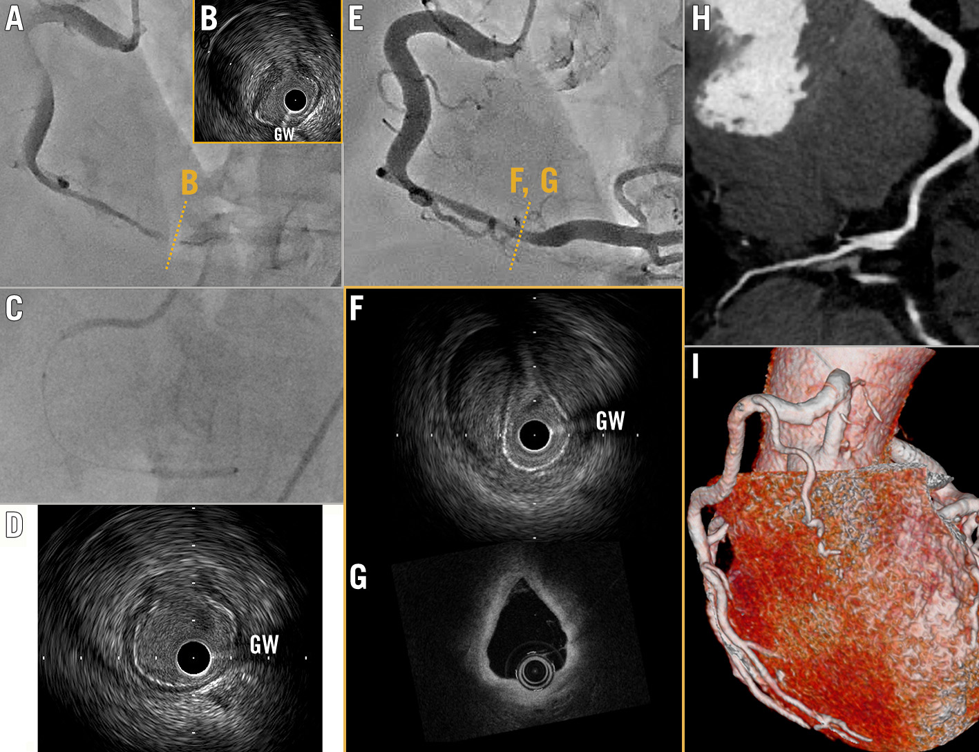
Figure 1. Intimal flap sealing with perfusion balloon. A) Coronary angiography (CAG) reveals significant stenosis in the right coronary artery. B) Intravascular ultrasound (IVUS) shows massive intramural haematoma and flap. C) Long inflation with a 2.5 mm perfusion balloon. D) IVUS shows satisfactory acute gain was achieved. E) Follow-up CAG on the 14th day of hospitalisation. F) IVUS and (G) optical coherence tomography (OCT) show the healed flap. H,I) Coronary computed tomography shows completely healed lesion 3 months later. GW: guidewire artefact

Conflict of interest statement

The authors have no conflicts of interest to declare.

Share

Supplementary data

https://www.asiaintervention.org/wp-content/uploads/2023/03/AIJv9i1_Oba_video1.mp4
Moving image 1. Urgent coronary angiography (CAG) revealed severe stenosis with Thrombolysis and Myocardial Infarction (TIMI) flow grade 1 in the right coronary artery (RCA).


https://www.asiaintervention.org/wp-content/uploads/2023/03/AIJv9i1_Oba_video2.mp4
Moving image 2. Intravascular ultrasound image showing an intimal flap and massive intramural haematoma.


https://www.asiaintervention.org/wp-content/uploads/2023/03/AIJv9i1_Oba_video3.mp4
Moving image 3. Intravascular ultrasound image after dilatation with a 2.5 mm perfusion balloon.


https://www.asiaintervention.org/wp-content/uploads/2023/03/AIJv9i1_Oba_video4.mp4
Moving image 4. Follow-up intravascular ultrasound on the 14th day of hospitalisation.


https://www.asiaintervention.org/wp-content/uploads/2023/03/AIJv9i1_Oba_video5.mp4
Moving image 5. Follow-up optical coherence tomography on the 14th day of hospitalisation.


References

  • Seggewiss H, Gleichmann U, Fassbender D, Vogt J, Mannebach H, Minami K. Therapy for acute vascular complications in percutaneous transluminal coronary angioplasty with the autoperfusion balloon catheter. Eur Heart J 1992;13:1649-57

Volume 9 - Number 1

View full issue

Download this article
Authors
  • Hajime Shinohara
  • Hiroshi Funayama
  • Kazuomi Kario
  • Keisuke Narita
  • Yusuke Oba
AsiaIntervention
  • Readers
    • Archives
    • Subscribe to the newsletter
    • Contact us
  • About the journal
    • Overview
    • Editorial Board
    • Masthead
  • Services
    • Advertising in AsiaIntervention
    • Article reprints
    • Publication calendar
    • Rights & Permissions
  • Authors
    • Authors guidelines
    • Submit your paper
  • Legal
    • Disclaimer
    • Cookies Policy
    • Privacy Policy
    • Legal Notice
  • Follow us
    • Facebook
    • Twitter
    • LinkedIn
Online ISSN 2491-0929 - Print ISSN 2426-3958
© 2015-2023 Europa Group - All rights reserved