Skip to content
The reference academic journal by and for the Asia-Pacific interventional cardiology community
AsiaIntervention

AsiaIntervention

  • Current Issue
  • Archives
  • How to submit
    • Authors guidelines
    • Submit your paper
  • Services
    • Advertising
    • Article reprints
    • Publication calendar
    • Rights & Permissions
  • About the journal
    • Overview
    • Editorial Board
    • Masthead
  • Contact us
Volume 6 – Number 2 – December 2020

Direct stenting disaster bailed out by intravascular lithotripsy

AsiaIntervention 2020;6:102-103 | 10.4244/AIJ-D-20-00001

Sandeep Basavarajaiah1, MD, MRCP, FESC; Kaeng Lee1, MD, MRCP; Gurbir Bhatia1, MD, MRCP; Michael Pitt1, MD, MRCP

1. Heartlands Hospital, University Hospitals Birmingham, Birmingham, United Kingdom

Sandeep Basavarajaiah

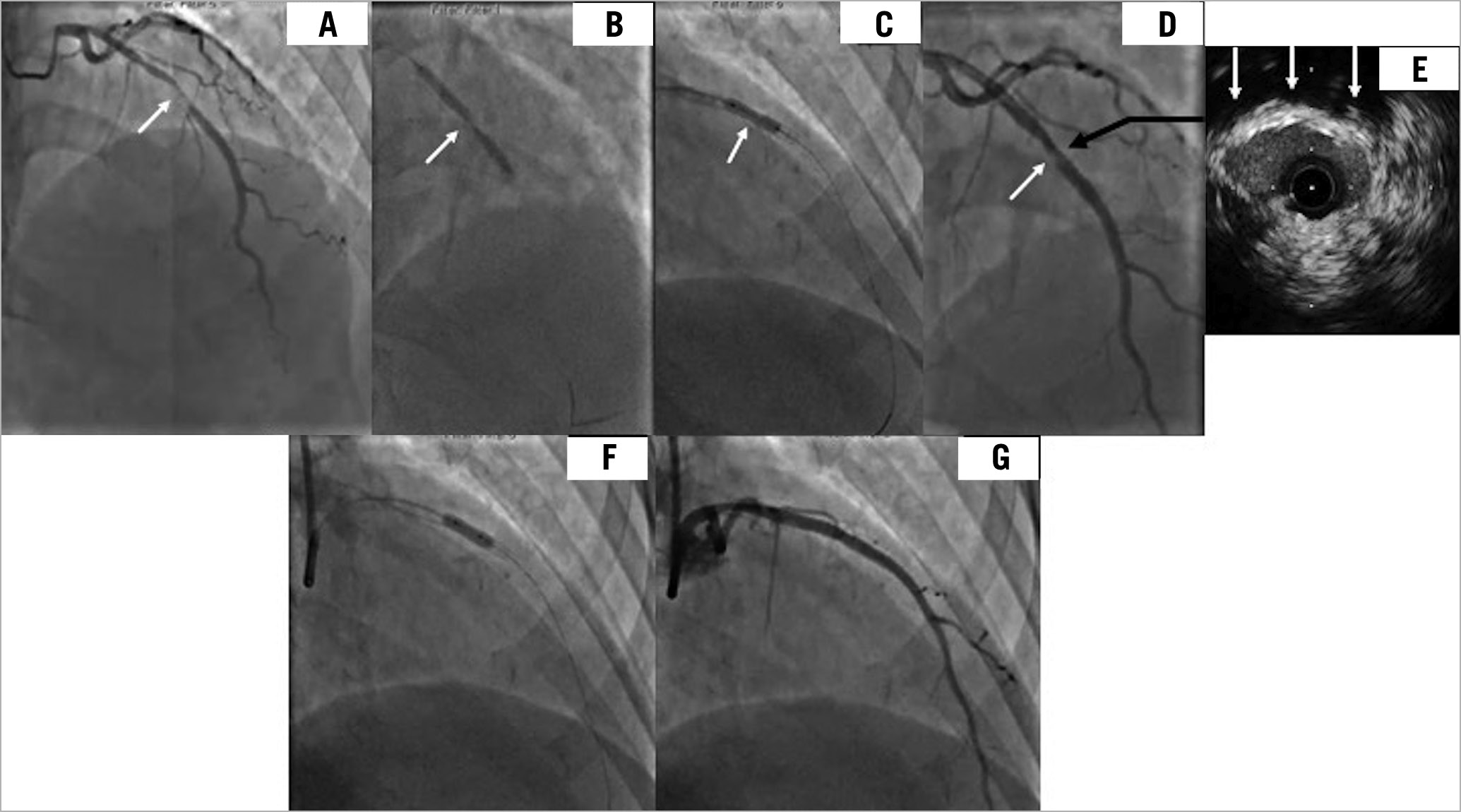
A 58-year-old woman with acute coronary syndrome had a significant lesion in the mid-segment of the LAD, which appeared smooth on the angiogram (Panel A, Moving image 1). The operator directly stented the lesion (3.0×38 mm DES) without pre-dilatation, which resulted in a focal area of significant under-expansion (Panel A & Panel B), and which failed to expand despite using a 3.0 high-pressure non-compliant balloon inflated to 35 atm (Panel C & Panel D). IVUS demonstrated the fibro-calcific nature of the lesion that resisted stent expansion and the minimum stent area was 3.5 mm2. (Panel A-Panel E & Moving image 2) Although, intravascular lithotripsy (IVL) is used before stent deployment in calcified undilatable lesions, there are no reports of its use in a recently deployed stent. We felt it was relatively safe to use IVL instead of larger-diameter high-pressure balloons that risked perforation. A 3.0 mm IVL balloon adequately expanded the stent (Panel F) and achieved a good final result (Panel G & Moving image 3). The patient was discharged 24 hours later and she remains free of any clinical events at 12 months. Although there appears to be a theoretical risk of polymer and drug disruption with IVL in a recently deployed stent, in our case, we felt that it was a safer option than high pressure balloons, and we have not seen any adverse clinical outcomes at 12 months. This case reinforces the message that no lesion should be stented directly without pre-dilatation, even if it appears smooth and non-calcific on the angiogram.

Conflict of interest statement

The authors have no conflicts of interest to declare.

Share

Supplementary data

https://www.asiaintervention.org/wp-content/uploads/2021/07/AIJv6i2_Basavarajaiah_video1.mp4

Moving image 1. Coronary angiogram demonstrating significant disease in the mid-segment of the LAD.

https://www.asiaintervention.org/wp-content/uploads/2021/07/AIJv6i2_Basavarajaiah_video2.mp4

Moving image 2. IVUS showing fibro-calcific lesion at the site of the under-expanded stent.

https://www.asiaintervention.org/wp-content/uploads/2021/07/AIJv6i2_Basavarajaiah_video3.mp4

Moving image 3. Final angiogram with improved stent expansion.

Volume 6 - Number 2

View full issue

Download this article
Authors
  • Gurbir Bhatia
  • Kaeng Lee
  • Michael Pitt
  • Sandeep Basavarajaiah
AsiaIntervention
  • Readers
    • Archives
    • Subscribe to the newsletter
    • Contact us
  • About the journal
    • Overview
    • Editorial Board
    • Masthead
  • Services
    • Advertising in AsiaIntervention
    • Article reprints
    • Publication calendar
    • Rights & Permissions
  • Authors
    • Authors guidelines
    • Submit your paper
  • Legal
    • Disclaimer
    • Cookies Policy
    • Privacy Policy
    • Legal Notice
  • Follow us
    • Facebook
    • Twitter
    • LinkedIn
Online ISSN 2491-0929 - Print ISSN 2426-3958
© 2015-2023 Europa Group - All rights reserved