Gunasekaran Sengottuvelu1*, MD, DM, FSCAI, FRCP; Ravindran Rajendran1,2, MD, DM; Raghul Ganesapandi1, MD, DM
AsiaIntervention 2019;5:130-131, DOI: 10.4244/AIJ-D-18-00032
1. Apollo Hospitals, Chennai, Tamil Nadu, India; 2. Trichy SRM Medical College Hospital & Research Centre, Irungalur Village, Tamil Nadu, India
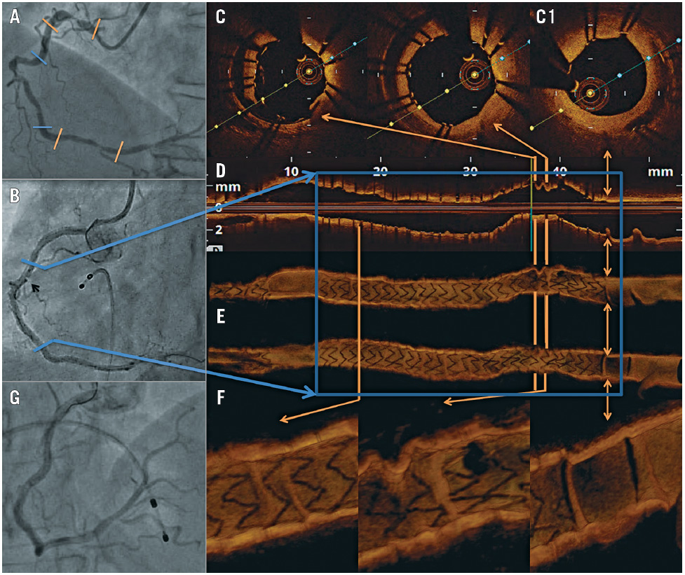
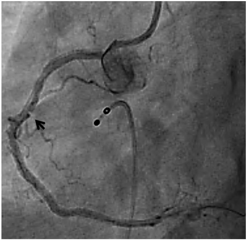
A 59-year-old male with effort angina was found to have tandem lesions involving the tortuous right coronary artery (RCA) (Panel A, Moving image 1). These were treated with three nonoverlapping drug-eluting stents in the distal, mid and ostio-proximal RCA, in that order. After deploying the long second stent across the tortuous mid RCA, an angiogram showed a distinct cut-like intraluminal filling defect within the proximal part of the stented segment, possibly a pseudostenosis due to straightening of the vessel and non-conformability (Panel B, Moving image 1).
Optical coherence tomography (OCT) imaging showed a well apposed stent with an area appearing like a cavity/dissection flap seen on the outer side of the stent struts which was actually infolding of the vessel wall due to the straightening of the tortuous segment by the stent (Panel C). OCT also showed infolding of the vessel proximal to the stent (not obvious on the angiogram) which appeared like a cavity with ruptured plaque (Panel C1). In the longitudinal view these areas were clearly seen as infolding of the vessels (Panel D). Once the wire was removed, there was only a mild irregularity seen within the proximal part of the mid RCA stent (Panel G, Moving image 1c). 3D OCT images revealed newer insights which were diagnostic of a concertina effect showing folds of vessel wall protruding through the stent struts that corresponded to the angiographic haziness and the dissection flaps on the axial OCT (Panel E, Panel F). In addition, there were multiple vessel wall infoldings all along the mid RCA stent that were not obvious on the angiogram, axial and longitudinal OCT. The straightening of tortuous coronary arteries by intracoronary guidewires has been well described as a concertina effect causing angiographic pseudostenosis and has been imaged using OCT. We report an unusual case of a concertina effect within and proximal to the stent caused by stenting of a tortuous vessel. 3D OCT images showed unique findings confirmative of a concertina effect and appear to be more sensitive than axial and longitudinal OCT. New lesions appearing during percutaneous coronary interventions are challenging to the operator and recognition of pseudostenosis is critical to avoid unnecessary stents. The axial OCT dissection-like appearance is due to the oblique cutting of the eccentric infolding by the arc of light. When these axial frames align and merge, it reforms the infolding of the vessel with smooth intima on the en face view in the 3D OCT (Supplementary Figure 1) A magnified view of the angiogram to show the concertina within the mid RCA stent is presented in Supplementary Figure 2.
Conflict of interest statement
The authors have no conflicts of interest to declare.
Supplementary data
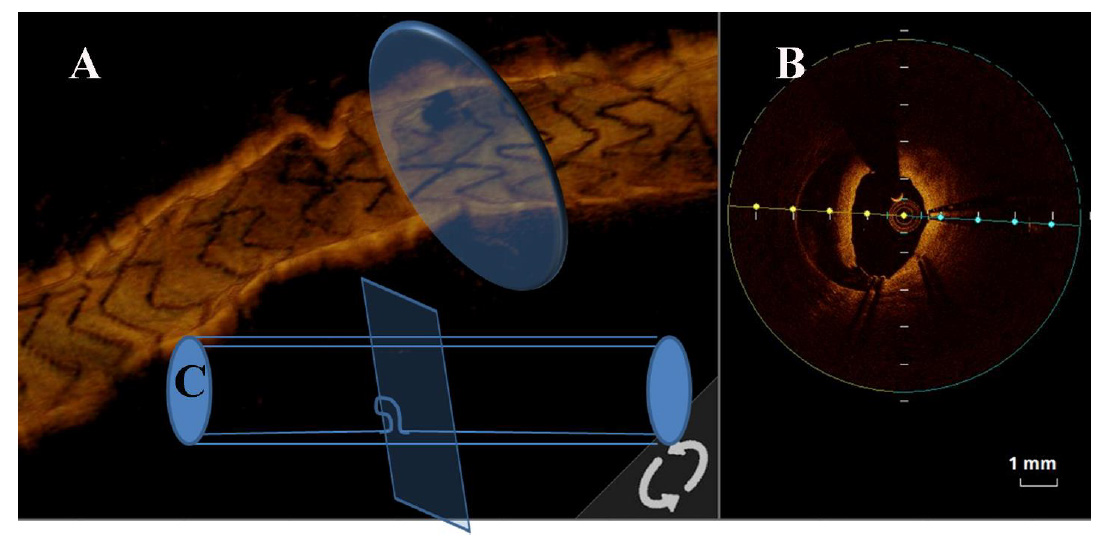
Supplementary Figure 1. Illustration of axial OCT having a dissection- like appearance (concertina effect).
A) 3D longitudinal OCT obliquely cut by an arc of light across the infolding area to produce an axial OCT mimicking dissection flaps as in panel B.
C) A line diagram to illustrate the above fact.
Moving image 1. Angiogram and axial OCT run of concertina effect. a. Tortuous RCA with multiple stenoses. b. Haziness and pseudostenosis after mid RCA stent. c. Final angiogram after removing the wire. d. Axial OCT run showing mimicking dissection flaps and ruptured cavities.
To download, please click below.