Skip to content
The reference academic journal by and for the Asia-Pacific interventional cardiology community
AsiaIntervention

AsiaIntervention

  • Current issue
  • Archives
  • How to submit
    • Authors guidelines
    • Submit your paper
    • Reviewers guidelines
  • Services
    • Advertising
    • Article reprints
    • Publication calendar
    • Rights & Permissions
  • About the journal
    • Overview
    • Editorial Board
    • Masthead
  • Contact us
Volume 9 – Number 2

Calcification with a figure of eight appearance found in routine coronary angiography

AsiaIntervention 2023;9:170-171 | 10.4244/AIJ-D-23-00014

Ramanathan Velayutham1, MD; A. Shaheer Ahmed1, MD, DM; Saurav Banerjee1, MD

1. Department of Cardiology, Jawaharlal Institute of Postgraduate Medical Education and Research, Puducherry, India

A 65-year-old female with recent anterior wall myocardial infarction presented with exertional angina. The examination found a pansystolic murmur at apex. Transthoracic echocardiography showed hypokinesia of the anteroseptal region and mitral annular calcification with severe mitral regurgitation.

Coronary angiography showed triple vessel disease. Fluoroscopy in the left anterior oblique (LAO) view demonstrated calcifications with a horizontal figure of eight appearance (Figure 1).

This image illustrates the importance of understanding fluoroscopic anatomy. Drawing an oblique line in the middle of the fluoroscopic LAO view helps to differentiate the right- and left-sided cardiac chambers1. The lower semicircle of the figure 8 was formed by annular calcification of the tricuspid valve and mitral valve annulus, while the upper half was completed by calcium along the right coronary artery and left circumflex artery on the right and left sides, respectively. The patient was referred for surgical mitral valve replacement with coronary artery bypass grafting.

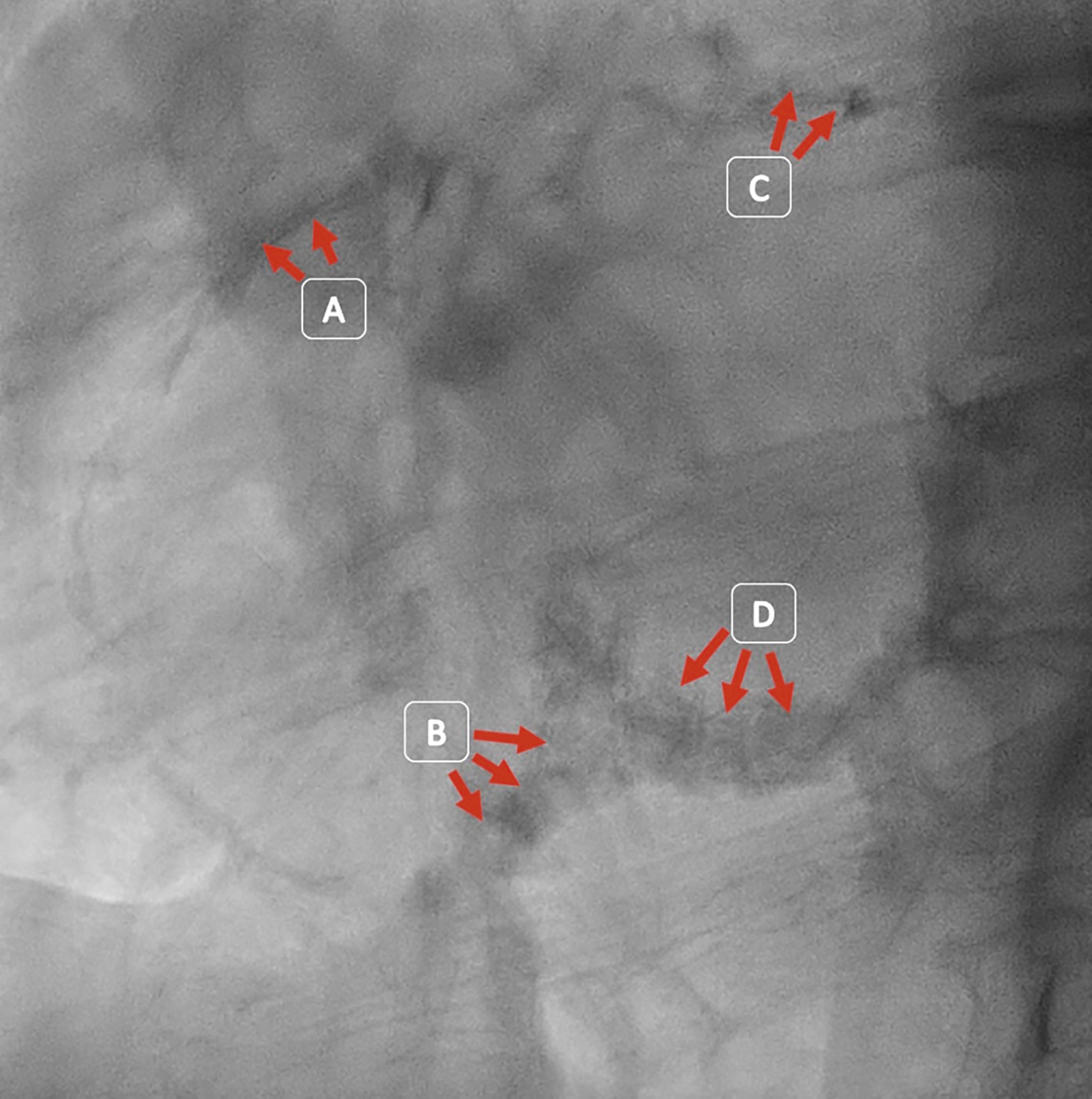
Figure 1. Fluoroscopic LAO view. This figure shows calcium deposits in a figure of eight along the right coronary artery (A), the tricuspid annulus (B), the left circumflex artery (C) and the mitral annulus (D). LAO: left anterior oblique

Conflict of interest statement

The authors have no conflicts of interest to declare.

Share

References

  • Thériault-Lauzier P, Andalib A, Martucci G, Mylotte D, Cecere R, Lange R, Tchétché D, Modine T, van Mieghem N, Windecker S, Buithieu J, Piazza N. Fluoroscopic anatomy of left-sided heart structures for transcatheter interventions: insight from multislice computed tomography. JACC Cardiovasc Interv 2014;7:947-57

Volume 9 – Number 2

View full issue

Download this article
Authors
  • A. Shaheer Ahmed
  • Ramanathan Velayutham
  • Saurav Banerjee
AsiaIntervention
  • Readers
    • Archives
    • Subscribe to the newsletter
    • Contact us
  • About the journal
    • Overview
    • Editorial Board
    • Masthead
  • Services
    • Advertising in AsiaIntervention
    • Article reprints
    • Publication calendar
    • Rights & Permissions
  • Authors
    • Authors guidelines
    • Submit your paper
  • Legal
    • Disclaimer
    • Cookies Policy
    • Privacy Policy
    • Legal Notice
  • Follow us
    • Facebook
    • X
    • LinkedIn
Online ISSN 2491-0929 - Print ISSN 2426-3958
© 2015-2025 Europa Group - All rights reserved