A 52-year-old female with hypertension and diabetes presented with acute onset chest pain. Electrocardiography showed a sinus rhythm with T-wave inversions in V1-V4, ST-segment depression in II, III, aVF, and V4-V6. The troponin T level was elevated. Echocardiography showed hypokinesia of the inferior wall with an ejection fraction of 45-50%. The patient was managed medically as a case of non-ST-segment elevation acute coronary syndrome. The next day, a coronary angiogram was done using a 6 Fr TIG (Terumo) catheter. An initial attempt was made to engage the left coronary artery, but it could not be visualised. While trying to engage the right coronary artery (RCA), the catheter entered a large branch arising from the superior aspect of the right sinus and supplying the left anterior descending artery (LAD) territory. The artery bifurcated within the anterior interventricular groove, with one branch coursing downwards, supplying the distal LAD territory, and the other branch coursing upwards, supplying the proximal LAD territory (Figure 1A, Figure 1B, Moving image 1, Moving image 2). This artery showed 70-80% disease in the proximal segment and diffuse disease distally. Angiograms were taken in different views. Initially, we suspected a possible left main total occlusion with collateral from the right side (conus branch engaged) or a possible coronary anomaly. Next, a right coronary artery injection was done, which showed a hyperdominant RCA with 90-95% mid-segment obstruction and diffuse disease in the right posterior descending artery (Figure 1C). The RCA was also seen supplying the left circumflex (LCx) territory through a large circumflex branch. Finally, an aortic root pigtail angiogram was performed, which showed no coronaries arising from the left sinus and a large right sinus (Figure 1D). Subsequently, a coronary computed tomography angiogram revealed the LAD and RCA both arising from the right sinus, with the LAD having a prepulmonary course (Figure 1E–Figure 1F–Figure 1G–Figure 1H, Moving image 3). Next, the patient successfully underwent percutaneous coronary intervention to the anomalous LAD and RCA.
The intervention for the anomalous LAD was challenging. Initially, a Judkins Right guide catheter was utilised; however, the balloon could not be tracked because of poor guide support. Next, an Amplatz Left 1 guide catheter was used to engage the anomalous LAD. In addition, two Runthrough NS Floppy coronary wires (Terumo) were used. One wire was positioned in the upper branch and a second wire in the lower branch of the LAD where it bifurcated in the interventricular groove. This facilitated better tracking of hardware and enabled successful intervention of the anomalous LAD. The patient was discharged the next day. She was followed up in the outpatient clinic one month after discharge and was asymptomatic.
Coronary artery anomalies are rare and are observed in 0.5-2% of the general population1. Though rare, they carry a high risk of mortality and morbidity when present. An anomalous origin of the left coronary artery arising from the right sinus of Valsalva is an uncommon malformation, accounting for 0.15% of cases1, and has been associated with myocardial ischaemia and sudden cardiac death2. Depending on the anatomical relationship of the anomalous vessel to the aorta and the pulmonary trunk, the anomaly can be classified into four common courses: posterior, interarterial, anterior, and septal. This patient had a prepulmonary course. This specific anomaly is highly uncommon3. Limaye et al3 presented a case report of a 50-year-old male with an inferior myocardial infarction who had undergone thrombolysis. Coronary angiography revealed total occlusion of the RCA and a coronary anomaly with separate origins of the LAD and LCx arteries from the right coronary sinus. Computed tomography showed a retroaortic LCx course and a prepulmonic LAD course, as in our case. Also, in our case, the LAD, arising from the right sinus, was seen dividing into a superior branch that supplies areas normally supplied by the proximal LAD and an inferior branch which supplies areas of the distal LAD. This specific anatomy of an anomalous LAD is rare. The prepulmonic course of the LAD is considered benign compared with the interarterial courses between the aorta and pulmonary artery, which are linked to adverse events, including sudden cardiac death4.
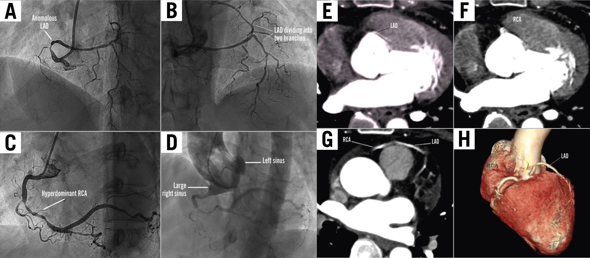
Figure 1. Coronary angiograms. A) LAO cranial view showing the anomalous LAD from the right sinus, with 70-80% disease in the proximal segment and diffuse disease distally. B) AP cranial view showing the anomalous LAD dividing into two branches within the interventricular groove. C) LAO cranial view showing the hyperdominant RCA with 90-95% mid-segment obstruction and diffuse disease in the RPDA. D) Pigtail angiogram of the aortic root in the AP view showing a large right sinus, with no coronaries arising from the left sinus. Coronary CT angiograms: E) Anomalous origin of the LAD from the right sinus. F) RCA arising from the right sinus. G) Distal courses of the LAD and the RCA. H) 3D reconstruction showing the anomalous LAD from the right sinus, with no coronaries arising from the left sinus. 3D: three-dimensional; AP: anterior-posterior; CT: computed tomography; LAD: left anterior descending artery; LAO: left anterior oblique; RCA: right coronary artery; RPDA: right posterior descending artery
Conflict of interest statement
The authors have no conflicts of interest to declare.