Skip to content
The reference academic journal by and for the Asia-Pacific interventional cardiology community
AsiaIntervention

AsiaIntervention

  • Current issue
  • Archives
  • How to submit
    • Authors guidelines
    • Submit your paper
    • Reviewers guidelines
  • Services
    • Advertising
    • Article reprints
    • Publication calendar
    • Rights & Permissions
  • About the journal
    • Overview
    • Editorial Board
    • Masthead
  • Contact us
Volume 9 – Number 2

Paracentral MitraClip implantation technique in a mitral valve with a small area due to rheumatic change

AsiaIntervention 2023;9:166-167 | 10.4244/AIJ-D-22-00063

Kento Kito1, MD; Akihisa Kataoka1, MD, PhD; Hirofumi Hioki1, MD, PhD; Yusuke Watanabe1, MD, PhD; Ken Kozuma1, MD, PhD

1. Department of Medicine, Teikyo University School of Medicine, Tokyo, Japan

An 85-year-old woman was referred to our hospital with severely symptomatic mitral regurgitation (MR). Transoesophageal echocardiography (TOE) showed a small mitral valve (MV) area (MVA) (2.21 cm2 on three-dimensional multiplanar reconstruction); mean MV pressure gradient (MVPG) of 1 mmHg, with restricted anterior mitral leaflet (AML) motion due to rheumatic changes, and MR jet from the A2/P2 region (Figure 1A–Figure 1C, Moving image 1, Moving image 2). Despite the rheumatic MV anatomy, our Heart Team offered transcatheter edge-to-edge repair because of the patient’s frailty and high surgical risk.

The procedure was performed on day 6 after admission. The MitraClip G4 NT (Abbott) was selected, and full grasping of the A2/P2 central region demonstrated worsened MR with unacceptably elevated MVPG (14 mmHg) and reduced MVA (0.43+0.35 cm2) (Figure 1D–Figure 1F, Moving image 3, Moving image 4). The target region was changed to the medial area of A2/P2, and full grasping showed a significant reduction of MR from 4+ to 2+, with acceptable MVPG (4 mmHg) and MVA (1.11+0.27 cm2) (Figure 1G–Figure 1I, Moving image 5, Moving image 6). The MitraClip was finally implanted without any complications. Thereafter, the patient was discharged on postoperative day 6 with reduced dyspnoea. One month after the procedure, the transthoracic echocardiography showed no evidence of MV damage, and the MR was reduced to 1+ with mildly elevated MVMG (8 mmHg). The patient was in a good condition and had no heart failure exacerbations during the 6-month follow-up.

Although the latest generation of MitraClip G4 NT was used in this case, it made no difference to the treatment quality because the clip size was the same as that of former generations. Central MitraClip implantation in the A2/P2 region is favoured over the paracentral position to prevent clinically relevant mitral stenosis1. As the AML is longest in the centre, implantation here yields lower valve tension and greater stability. However, owing to the reduced AML flexibility in rheumatic MVs, central implantation may unacceptably increase the MV tension, resulting in failed MR reduction and elevated MVPG due to significant residual MR, together with smaller neo-MVA. In contrast, A2/P2 paracentral implantation may stabilise the leaflet coaptation without excessively increasing the valve tension and MVPG, as it preserves the lateral AML motion. This technique provides a larger MVA and more MR reduction than central implantation. While a rheumatic MV anatomy is generally considered a relative contraindication, the paracentral implantation technique may be useful to avoid clinically significant mitral stenosis when a MitraClip is necessary.

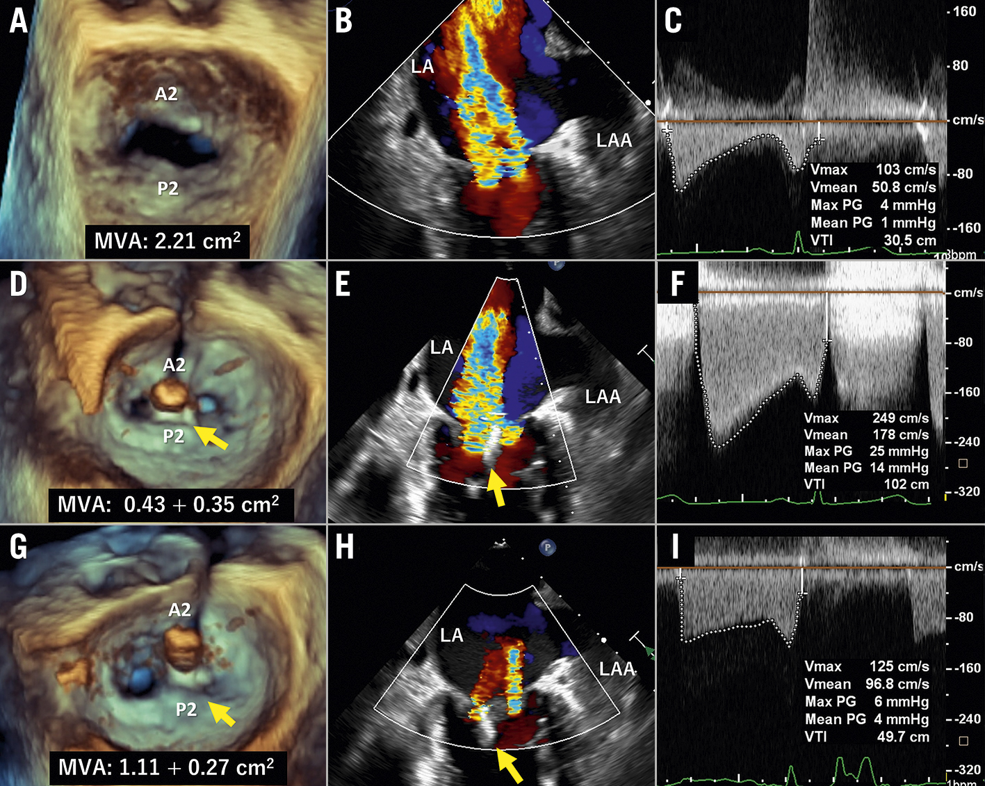
Figure 1. Transoesophageal echocardiography images at screening and during the procedure. En face commissure, and mitral valve pressure gradient images, at screening (A-C) and during the procedure after the central (D-F) and paracentral full grasping (G-I). Yellow arrows indicate the MitraClip. LA: left atrium; LAA: left atrial appendage; MVA: mitral valve area

Conflict of interest statement

A. Kataoka and Y. Watanabe received remuneration from Abbott Medical Japan as proctors for MitraClip. The other authors have no conflicts of interest to declare.

Share

Supplementary data

https://www.asiaintervention.org/wp-content/uploads/2023/09/AIJv9i2_Kito_video1.mp4
Moving image 1. 3D-TOE video of the en face view before the procedure shows a small mitral valve area (2.21 cm2 measured by 3D-MPR) and restrictive mitral valve leaflet motion. (corresponds to Figure 1A).


https://www.asiaintervention.org/wp-content/uploads/2023/09/AIJv9i2_Kito_video2.mp4

Moving image 2. 2D-TOE colour video of the commissure view before the procedure shows severe MR jet from A2/P2 region (corresponds to Figure 1B).


https://www.asiaintervention.org/wp-content/uploads/2023/09/AIJv9i2_Kito_video3.mp4
Moving image 3. 3D-TOE video of the en face view after full grasping of the MitraClip NT to the A2/P2 central region shows strikingly decreased mitral valve area (0.43+0.26 cm2 measured by 3D-MPR) (corresponds to Figure 1D).


https://www.asiaintervention.org/wp-content/uploads/2023/09/AIJv9i2_Kito_video4.mp4

Moving image 4. 2D-TOE colour video of the commissure view after full grasping of the MitraClip NT to the A2/P2 central region shows severe split MR jets (corresponds to Figure 1E).


https://www.asiaintervention.org/wp-content/uploads/2023/09/AIJv9i2_Kito_video5.mp4

Moving image 5. 3D-TOE video of the en face view after full grasping of the MitraClip NT to the A2/P2 medial region (paracentral) shows greater mitral valve area (1.11+0.27 cm2 measured by 3D-MPR) than that of the A2/P2 central region grasping (corresponds to Figure 1G).


https://www.asiaintervention.org/wp-content/uploads/2023/09/AIJv9i2_Kito_video6.mp4

Moving image 6. 2D-TOE colour video of the commissure view after full grasping of the MitraClip NT to the A2/P2 medial region shows decreased MR jets from 4+ to 2+ (corresponds to Figure 1H).


References

  • Kassar M, Praz F, Hunziker L, Pilgrim T, Windecker S, Seiler C, Brugger N. Anatomical and Technical Predictors of Three-Dimensional Mitral Valve Area Reduction After Transcatheter Edge-To-Edge Repair. J Am Soc Echocardiogr 2022;35:96-104

Volume 9 – Number 2

View full issue

Download this article
Authors
  • Akihisa Kataoka
  • Hirofumi Hioki
  • Ken Kozuma
  • Kento Kito
  • Yusuke Watanabe
AsiaIntervention
  • Readers
    • Archives
    • Subscribe to the newsletter
    • Contact us
  • About the journal
    • Overview
    • Editorial Board
    • Masthead
  • Services
    • Advertising in AsiaIntervention
    • Article reprints
    • Publication calendar
    • Rights & Permissions
  • Authors
    • Authors guidelines
    • Submit your paper
  • Legal
    • Disclaimer
    • Cookies Policy
    • Privacy Policy
    • Legal Notice
  • Follow us
    • Facebook
    • X
    • LinkedIn
Online ISSN 2491-0929 - Print ISSN 2426-3958
© 2015-2025 Europa Group - All rights reserved