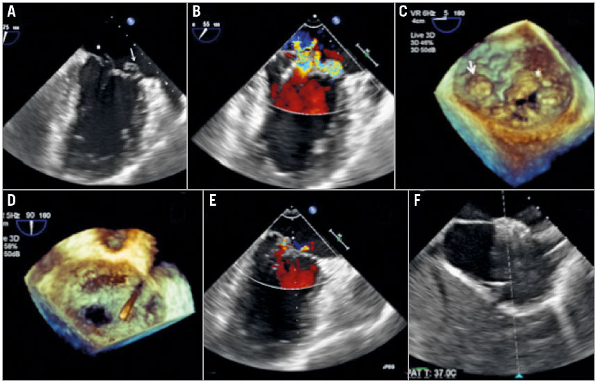
Figure 1. Transoesophageal echocardiography images during the MitraClip procedure.
Figure 1. Transoesophageal echocardiography images during the MitraClip procedure. A) Bi-commissural view of the transoesophageal echocardiogram demonstrating the complexity of the Barlow’s mitral valve. Arrow marks the medial A3 flail segment and the asterisk marks the P1 prolapse segment of the mitral valve. B) Bi-commissural view with colour Doppler demonstrating the significant mitral regurgitation arising from both the lateral and the medial site of the mitral valve. C) Three-dimensional en face view elucidating the complexity of the Barlow’s mitral valve. Arrow marks the medial A3 flail segment and the asterisk marks the P1 prolapse segment of the mitral valve. D) Three-dimensional en face view demonstrating a total of three clips successfully implanted. E) Bi-commissural view of the transoesophageal echocardiogram showing only trivial mitral regurgitation after the procedure. F) The iatrogenic atrial septal defect was closed by an AMPLATZER atrial septal defect occluder.