Chee Yang Chin, Calvin Woon Loong Chin, et al
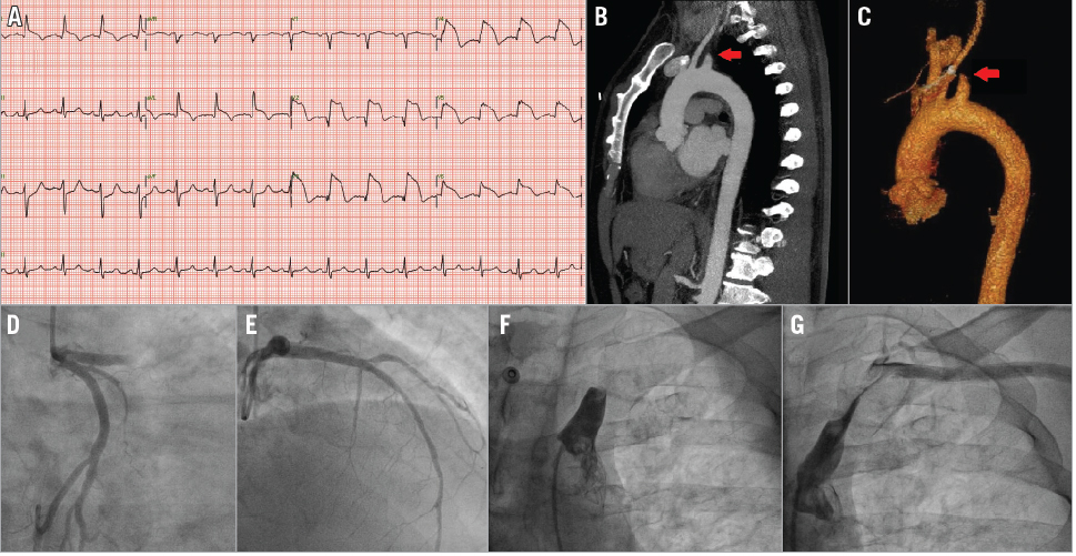
Simultaneous dual territory ischaemia is uncommon but, if misdiagnosed, can lead to delay in appropriate treatment. A middle-aged hypertensive male presented with acute chest pain and simultaneous left hand discolouration and coldness. Electrocardiography showed anterolateral ST-elevation (Panel A). Left upper limb examination revealed absent pulses, systolic blood pressure 50 mmHg less than the right and a cyanosed hand. An urgent CT aortogram excluded aortic dissection and revealed acute proximal left subclavian artery (LSA) occlusion (Panel B, Panel C). Successful primary PCI was performed to the proximal left anterior descending artery (Panel D, Panel E, Moving image 1, Moving image 2). LSA angiography confirmed proximal thrombosis (Panel F, Moving image 3), and aspiration thrombectomy achieved partial re-canalisation (Panel G, Moving image 4). Limb ischaemia steadily improved and surgery was deferred. Echocardiography and cardiac MRI did not reveal intracardiac thrombus. The patient was discharged on warfarin and dual antiplatelet therapy. Six weeks later, an ultrasound scan showed LSA patency with 50% residual stenosis.
Acute upper limb ischaemia with STEMI necessitates urgent exclusion of aortic dissection, which mandates completely different management. Subclavian artery thrombosis causing simultaneous acute myocardial infarction is uncommon and previously reported only in patients after coronary bypass grafting with the internal mammary artery. In our case, simultaneous dual vessel embolism or simultaneous plaque rupture, although rare, could explain the double occlusion. More likely, the patient had suffered an acute STEMI leading to rapid intracardiac clot formation and embolisation to the LSA. Early cardioembolism following MI is a recognised phenomenon and must be considered in patients presenting with simultaneous acute MI and non-coronary ischaemia.
Supplementary data
[/custom_font]
Moving image 1. Coronary angiography showing acute occlusion of the proximal LAD.
Moving image 2. Coronary angiography after percutaneous coronary intervention and stenting of the LAD.
Moving image 3. Angiography showing acute occlusion of the proximal left subclavian artery (LSA).
Moving image 4. Angiography after aspiration thrombectomy of the LSA.