Skip to content
The reference academic journal by and for the Asia-Pacific interventional cardiology community
AsiaIntervention

AsiaIntervention

  • Current issue
  • Archives
  • How to submit
    • Authors guidelines
    • Submit your paper
    • Reviewers guidelines
  • Services
    • Advertising
    • Article reprints
    • Publication calendar
    • Rights & Permissions
  • About the journal
    • Overview
    • Editorial Board
    • Masthead
  • Contact us
Volume 7 – Number 1 – July 2021

Pseudopericardial effusion due to excess tension caused by a guide catheter during the MitraClip procedure

AsiaIntervention 2021;7:60-61 | 10.4244/AIJ-D-21-00006

Ruri Ishibashi1, MD; Akihisa Kataoka1, MD, PhD; Yusuke Watanabe1, MD, PhD; Ken Kozuma1, MD, PhD

1. Department of Medicine, Division of Cardiology, Teikyo University School of Medicine, Tokyo, Japan

Pseudopericardial effusion due to excess tension caused by a guide catheter during the MitraClip procedure

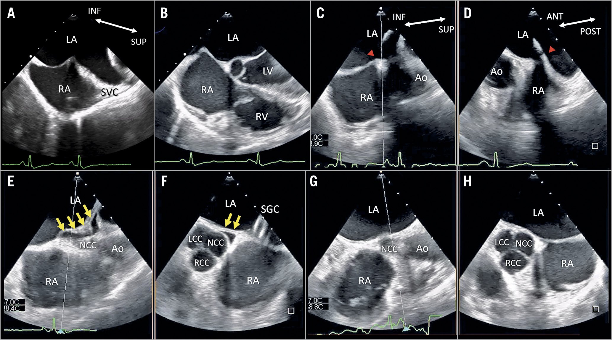
A 66-year-old man underwent transcatheter mitral valve repair for severe functional mitral regurgitation (MR) due to dilative cardiomyopathy.

There was no pericardial effusion (PE) (Panel A, Panel B, Moving image 1, Moving image 2), and transseptal puncture (TSP) was performed in the superior and anterior fossa ovalis without complications. Transoesophageal echocardiography (TEE) (EPIQ7/X8-2t system; Philips, Andover, MA, USA) revealed that the steerable guide catheter (SGC) was pulling the osseous membrane and causing it to tent as it was inserted into the left atrial (LA) cavity (Panel C, Panel D, Moving image 3). A MitraClip G4 NTW® (Abbott Vascular, Santa Clara, CA, USA) was successfully implanted once the MR had reduced. When the clip delivery system was removed from the SGC, TEE revealed a pericardial space between the LA wall and aortic root, indicating PE) (Panel E, Panel F, Moving image 4). No PE was observed elsewhere, and the vital signs remained unchanged, thereby excluding cardiac tamponade. The procedure was immediately terminated. However, the PE disappeared once the SGC was removed and did not return (Panel G, Panel H, Moving image 5, Moving image 6).

PE between the LA wall and aortic root is common. This fatal complication of TSP stems from improper puncture of the fossa ovalis1. However, we detected no such punctures using TEE. There is a pericardial sac, which has a double-walled structure, surrounded by the LA, right atrium, and base of the aortic root. We suspected that the redundant LA wall and pericardium were pulled due to excess tension generated by the SGC and displaced in the direction of the LA cavity, thereby creating free space. The Heart Team should consider pseudo-PE when excess tension is generated by the SGC during the MitraClip procedure.

Conflict of interest statement

The authors have no conflicts of interest to declare.

Share

Supplementary data

 
https://www.asiaintervention.org/wp-content/uploads/2021/07/AIJv7i1_Ishibashi_video1.mp4
Moving image 1. Bi-caval, four-chamber, and live xPlane images (bi-caval and short-axis views) of TEE performed during the MitraClip procedure. Transseptal puncture was performed in the superior and anterior fossa ovalis without complications.


https://www.asiaintervention.org/wp-content/uploads/2021/07/AIJv7i1_Ishibashi_video2.mp4
Moving image 2. Bi-caval, four-chamber, and live xPlane images (bi-caval and short-axis views) of TEE performed during the MitraClip procedure. Transseptal puncture was performed in the superior and anterior fossa ovalis without complications.


https://www.asiaintervention.org/wp-content/uploads/2021/07/AIJv7i1_Ishibashi_video3.mp4
Moving image 3. At the insertion of the SGC, the tenting of the intraseptal wall is shown.


https://www.asiaintervention.org/wp-content/uploads/2021/07/AIJv7i1_Ishibashi_video4.mp4
Moving image 4. After the removal of the clip delivery system, the pericardial effusion between the LA wall and aortic root is shown.


https://www.asiaintervention.org/wp-content/uploads/2021/07/AIJv7i1_Ishibashi_video5.mp4
Moving image 5. After the removal of the SGC, the pericardial effusion immediately disappeared.


https://www.asiaintervention.org/wp-content/uploads/2021/07/AIJv7i1_Ishibashi_video6.mp4
Moving image 6. After the removal of the SGC, the pericardial effusion immediately disappeared.

References

  • Salghetti F, Sieira J, Chierchia GB, Curnis A, de Asmundis C. Recognizing and reacting to complications of trans-septal puncture. Expert Rev Cardiovasc Ther 2017;15:905-12

Volume 7 - Number 1

View full issue

Download this article
Authors
  • Akihisa Kataoka
  • Ken Kozuma
  • Ruri Ishibashi
  • Yusuke Watanabe
AsiaIntervention
  • Readers
    • Archives
    • Subscribe to the newsletter
    • Contact us
  • About the journal
    • Overview
    • Editorial Board
    • Masthead
  • Services
    • Advertising in AsiaIntervention
    • Article reprints
    • Publication calendar
    • Rights & Permissions
  • Authors
    • Authors guidelines
    • Submit your paper
  • Legal
    • Disclaimer
    • Cookies Policy
    • Privacy Policy
    • Legal Notice
  • Follow us
    • Facebook
    • X
    • LinkedIn
Online ISSN 2491-0929 - Print ISSN 2426-3958
© 2015-2025 Europa Group - All rights reserved