An 80-year-old woman presented with dyspnoea. Coronary angiography revealed stenosis of the left main trunk (LMT) and the left anterior descending artery (LAD). The fractional flow reserve (FFR) was 0.64, which led to a percutaneous coronary intervention. After debulking the calcified nodules in the LMT using an orbital atherectomy system, optical coherence tomography (OCT) was performed (Figure 1A). Figure 1B illustrates the obtained image. We reviewed the three-dimensional (3D) reconstruction image, which revealed a bent OCT catheter (Dragonfly OPTIS [Abbott]) (Figure 1C). Following balloon dilation, the FFR improved to 0.89. We performed dilation with a drug-coated balloon between the LMT and LAD and ensured that the procedure was carried out with no complications. After the procedure, we found that the OCT catheter had broken between the lens and the tip, forming a Z shape (Figure 1D).
Two case reports described the Z-shape phenomenon using OCT as a potential risk factor for coronary injury12. In our study, we obtained OCT images with a “three circles sign” (Figure 1B), similar to those reported by Ha et al1. We investigated whether this image corresponded to that reported by Warisawa et al2 using a vascular model. We employed both the older (Dragonfly OPTIS) and newer versions of the OCT catheter (Dragonfly Opstar [Abbott]). When the catheter tip was secured and advanced in vitro, the OPTIS consistently replicated the Z-shape phenomenon (Figure 1E), and the OCT images were consistent with the previously observed images (Figure 1F). A 3D reconstruction image similar to Figure 1C was also obtained (Figure 1G). During endoscopy, the OCT catheter was flexed in the same position, as shown in Figure 1D, forming a Z shape and making contact with the vessel wall (Figure 1H), providing evidence of potential vessel damage. In contrast, the Opstar failed to produce a Z shape under similar conditions, likely due to its stiffer shaft. Our study verified that the “three circles sign” and the “Z-shape phenomenon” are identical using the vascular model. The new Opstar OCT catheter is potentially effective in reducing the risk of vascular injury caused by the Z-shape phenomenon.
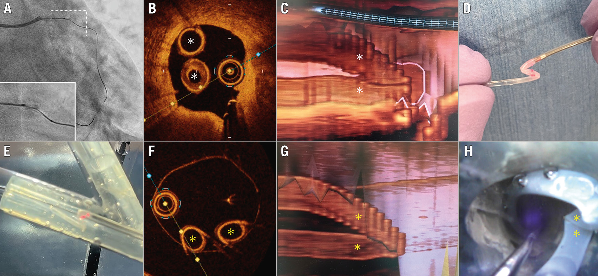
Figure 1. Optical coherence tomography (OCT) findings. A) OCT catheter inserted into the left anterior descending artery. The large white rectangle is a magnification of the small dotted one. The OCT catheter is bent. B) The cross-sectional OCT image displays the “three circles sign”. White asterisks indicate the bent OCT catheter shaft. C) Three-dimensional reconstruction reveals the Z-shaped OCT catheter. White asterisks highlight the bent OCT catheter shaft shown in B. D) The OCT catheter in the Z shape. E) Optis produces the Z shape after the catheter tip was secured and pushed inside the vascular model in vitro, similar to D. F) Cross-sectional OCT images captured during the process depicted in E. The image is similar to that in B. Yellow asterisks indicate the bent OCT catheter shaft. G) Three-dimensional image displays the Z-shaped OCT catheter. Yellow asterisks highlight the bent OCT catheter shaft depicted in F. H) Observation of the Z-shaped OCT catheter in the vascular model using endoscopy.
Conflict of interest statement
The authors have no conflicts of interest to declare.