Tatsuya Shiraki*, MD; Masaki Awata, MD, PhD; Keita Okayama, MD, PhD; Isamu Mizote, MD, PhD; Yasushi Sakata, MD, PhD
AsiaIntervention 2019;5:52, DOI: 10.4244/AIJ-D-18-00037
Department of Cardiovascular Medicine, Osaka University Graduate School of Medicine, Osaka, Japan
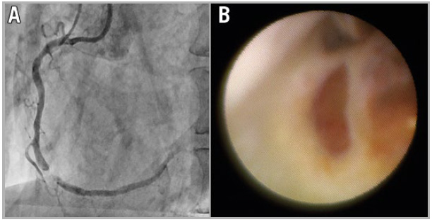
An 82-year-old male patient presented with a 99% stenosis in the middle part of the right coronary artery (Panel A). Percutaneous coronary intervention was performed with the latest angioscope (Forwardlooking®; Taisho Biomed Instruments Co., Ltd., Osaka, Japan). The angioscope is of the non-occlusion type, consisting of a probe catheter which has a monorail wire lumen and a fibre catheter and has a resolution of 9,000 pixels with full colour. After the culprit lesion was crossed with a 0.014-inch wire, the angioscopy catheter was advanced to the lesion with the Guide Plus™ guide extension catheter (NIPRO, Osaka, Japan) as delivery catheter and 10% dextran was continuously flushed through the delivery catheter for the displacement of blood at a rate of 4 mL/second for a total of 40 mL by a power injector. Then, all sequences were recorded using a digital recording system to observe the angioscopic movie. The lack of a fibrous cap and the exposed lipid core of the plaque were clearly recognised on angioscopy (Panel B, Moving image 1). The surface of the ruptured fibrous cap was yellow and some gloss findings were suggestive of the presence of cholesterol crystals. Based on this finding, distal protection was initiated and, subsequently, the stent was implanted. Although coronary slow-flow phenomenon had occurred, the coronary flow was immediately improved after removal of the distal protection catheter. Attenuated plaque by intravascular ultrasound and thincap fibroatheroma by optical coherence tomography have been reported as the cause of no-reflow phenomenon. As in this case, the ruptured fibrous cap seen on angioscopy may also be related to no-reflow phenomenon.
Conflict of interest statement
The authors have no conflicts of interest to declare.
Supplementary data
Moving image 1. The lack of a fibrous cap and the exposed lipid core of the plaque were clearly recognisable on angioscopy.
To download, please click below.