
X-rays, along with iodinated contrast media, are used to visualise cardiac arteries and chambers, which is a necessity for cardiac interventions. The visualisation of cardiac structures and arteries in real time is essential to facilitate percutaneous coronary and structural interventions. However, unlike plain film radiographs, fluoroscopy and digital acquisitions require multiple frames, which are taken in quick succession to produce moving images. As such, radiation usage and patient exposure are much higher than for plain film radiography. Furthermore, the duration and complexity of the procedure often expose patients and staff to higher radiation rates compared with other medical imaging studies.
Radiation exposure to the patient results from the interaction of X-rays with the patient’s tissues and organs. X-rays travel through the patient, reaching the detector and contributing to the production of an image. They are also absorbed by the patient, resulting in radiation exposure for the patient, or they are scattered off the patient’s tissues and into the surrounding environment, including the staff performing the procedure.
Current International Commission on Radiological Protection (ICRP) recommendations set an occupational effective dose limit of 20 mSv/year averaged over 5 years, with no single year >50 mSv. The lens limit is 20 mSv/year averaged over 5 years (no single year >50 mSv), while skin and extremity limits remain 500 mSv/year. The public limit is 1 mSv/year. Patients have no fixed numerical limit; medically indicated exposure is governed by justification, optimisation, and diagnostic reference levels (DRLs).12 These limits are summarised in Table 1.
Staff exposure is primarily from scatter and is associated with cataracts, cellular/chromosomal injury, and possible cancer risk.345 Prevention relies on minimising the cumulative dose, shielding, and dosimetric surveillance.
Unlike the hazards above, musculoskeletal injury in the cardiac catheterisation laboratory (CCL) arises from cumulative mechanical loading rather than from radiation itself. The principal cause is the 5-10 kg weight of a lead apron, borne asymmetrically over years of practice, combined with prolonged static standing and awkward cervical/spinal positioning while viewing overhead monitors. The most commonly reported injuries are lumbar disc disease, cervical spondylosis, and knee or hip osteoarthritis. Orthopaedic problems related to protective wear were the most frequently reported radiation-associated health hazard in the European Association of Percutaneous Cardiovascular Interventions (EAPCI) survey (19.5%), with an association noted with increasing years of exposure;6 furthermore, two-thirds of respondents to a 2024 European survey reported garment-related orthopaedic injury personally or in coworkers.7 In the Women-in-Asian Pacific Society of Interventional Cardiology (WIN-APSIC) survey, orthopaedic injury was the most commonly reported occupational health effect (13.4%), with lumbar disc injury the most frequent, yet only 32% of respondents had any exercise routine to mitigate this risk, and very few institutions offered structured support.8
Because these injuries are mechanical rather than radiobiological, prevention should be addressed separately from radiation-dose mitigation. Measures include weight-distributing two-piece aprons, lightweight or suspended protection systems, ergonomic positioning, strengthening and stretching programmes, scheduled breaks or operator rotation, and access to physiotherapy or occupational health review.
Table 1. Current radiation dose limits – occupational workers, the public, and patients.
| Population | Dose limit | Notes |
|---|---|---|
| Occupational workers (effective dose) | 20 mSv/year, averaged over 5 years; no single year >50 mSv | ICRP recommendation 1 |
| Occupational workers (lens of eye) | 20 mSv/year, averaged over 5 years; no single year >50 mSv (reduced from 150 mSv/year) | Since ICRP 2011 statement on tissue reactions 2 |
| Occupational workers (skin, extremities) | 500 mSv/year | ICRP recommendation 2 |
| Members of the public | 1 mSv/year | ICRP recommendation 1 |
| Patients (medical exposure) | No fixed numerical dose limit | Controlled by justification and optimisation (DRLs) rather than a dose limit, as each exposure is clinically indicated |
| Current radiation dose limits for occupational workers and members of the public and the optimisation-based approach used for medically indicated patient exposure. Patients do not have a fixed numerical dose limit; exposure is governed by clinical justification, optimisation, and DRLs. DRL: diagnostic reference level; ICRP: International Commission on Radiological Protection; mSv: millisievert | ||
Radiation protection techniques
Radiation reduction is based on minimising exposure time, maximising distance, and shielding. Patient dose can be reduced through lower frame rates, fewer and shorter cine runs, and efficient procedures (Table 2).9 Beam collimation, low pulse/frame rates, limited magnification and steep angulation, spectral filtration, optimised source-detector geometry, last-image hold, and avoidance of unnecessary cine runs reduce primary and scatter radiation.910
Dose area product (DAP), or kerma area product (KAP), combines dose and the area of the patient exposed and is widely used to compare procedures and establish DRLs. The American College of Cardiology (ACC) recommends investigation when a CCL’s median KAP exceeds the 75th percentile.11121314
The Asian Pacific Society of Interventional Cardiology (APSIC) provides an Asia-Pacific (APAC) perspective on radiation safety. Regional DRLs vary: Japanese studies report 59.3-64 Gy·cm² for diagnostic angiography,12 Australian data 30-59 Gy·cm²,1314, Thailand reports 91 Gy·cm² for percutaneous coronary intervention (PCI),15 and India 20 Gy·cm² and 50 Gy·cm² for diagnostic angiography and PCI, respectively.1617 A European study reported 35 Gy·cm² for diagnostic angiography and 85 Gy·cm² for PCI.18 No coordinated APAC-wide DRL survey has been completed; standardised regional reporting is needed to quantify variation and guide dose reduction.
Variation in radiation dose amongst APAC countries is noted, and the Asia-Pacific region may benefit from further coordinated reporting. Global variations in available resources, practice, and policy are noted. The WIN-APSIC radiation safety survey,8 evaluating radiation hazards and safety among physician and allied health care professionals in invasive cardiology, reveals significant international variability in radiation safety practices and training internationally and in the Asia-Pacific region. Further comparative DRL data from the Asia-Pacific region documenting exposure rates with comparative methodology are important to accurately quantify the impact of these differences and minimise exposure and risk.
Table 2. Measures of radiation exposure.
| Measure | Definition |
|---|---|
| Fluoroscopy time | The duration for which the main operator has the X-ray system’s fluoroscopy function activated. Fluoroscopy is a low-dose imaging mode used to position catheters and wires, though with lower image quality than digital acquisition, and images are not routinely stored. It indicates procedure complexity/length but does not measure dose. |
| Air kerma | This is the “kinetic energy released per mass unit of air”. It is an important measure that can be used to quantify the output of the X-ray system and the intensity of the exposure to the patient. It does not include the area of patient exposed. |
| Kerma area product | Also referred to as dose area product, it measures air kerma and the area of the patient exposed. It is the most useful radiation metric as it captures the total radiation used during the procedure and correlates to exposure to staff in close proximity. |
| Number of digital acquisitions | Also referred to as the number of “cine runs”. It dates back to when cinematography with film cameras was used in angiography. |
| Absorbed dose | This is the amount of energy deposited per unit mass of tissue and is useful to assess the biological risk to that tissue. It is reported in Gy or mGy. |
| Effective dose | This is a whole-body dose that assesses an individual’s stochastic risk, which is useful when comparing risks between exposed individuals. It is measured in Sv or mSv. |
| Exposure | This is the total charge of ions produced by radiation at a specific point. It assesses the level of risk at the specified location where energy is released. It is measured in Gy. |
| Equivalent dose | This is the dose quantity that factors in the relative biological damage and type of radiation used. It is used to measure risk for specific tissues for staff and is measured in Sv or mSv. |
| Summary of commonly used radiation exposure and dose metrics in the cardiac catheterisation laboratory, including what each measure represents and its principal unit or clinical interpretation. Gy: gray; mGy: milligray; mSv: millisievert; Sv: sievert | |
Global differences
Radiation practices and regulation vary substantially across APAC. In the United States, cardiology procedures account for approximately 45% of the mean cumulative medical radiation exposure, and interventional cardiologists historically have among the highest occupational exposures, although this difference is narrowing within the medical profession.19
In the 2012 EAPCI survey of 615 CCL workers, 64.4% always used a collar dosimeter, whereas 7.0% never did. Thyroid shields were used by 87.2%, lead glasses by 38.4%, and leg shields by 31.9%. Orthopaedic problems from protective garments were reported by 19.5%, cataracts by 2.0%, blood-count abnormalities by 2.4%, and malignancy by 2.2%. Most respondents (62.1%) reported workplace restrictions for pregnant workers.620
A more recent (2024) survey conducted among European countries (where the majority of respondents were electrophysiologists) found that 57.3% of respondents reported mandatory radiation training was required for clinical practice.7 Although a European directive on radiation use (Euratom 2013/59) exists, 54.5% reported being unfamiliar with this document. Two-thirds reported radiation protection garment-related orthopaedic injuries (personally or in coworkers) as a result of working with occupational radiation, with similar proportions not knowing if these were recognised as occupational hazards in their countries. The majority of respondents (78.7%) were unaware of the identity of their centre’s radiation protection officer.7
A European Association of Cardiovascular Imaging (EACVI) survey of 258 respondents showed substantial variability in protection for interventional echocardiographers (IEs): only 28% received periodic radioprotection training, 33% lacked dedicated architectural shielding, and 72% did not know their annual dose; 72% called for improved protection.21
The WIN-APSIC radiation safety survey (2024-2025),8 evaluating radiation hazards and safety among physician and allied health care professionals in invasive cardiology, reports important variation in practice. In this survey, 76.6% of respondents worked in APSIC member countries (72.84% interventional cardiologists, 3.97% electrophysiologists, and 25.2% allied health professionals). Survey respondents reported that 86.8% wore lead aprons and 83.2% wore thyroid collars, 30% wore lead glasses, 4.3% used shin/leg guards, and 13.33% of females reported the use of pauldron breast protection shielding (lead gowns fitted with a lead pauldron).15
WIN-APSIC also found limited use of advanced protection and dosimetry: 4.6% had Zero-Gravity systems (TIDI Products), 25% wore no dosimeter, 63.2% received dosimeter feedback, 55.9% used radiation dose as a CCL quality metric, and 57.7% had a radiation officer. Mandatory radiation-protection education was reported by 73.4%.8
Orthopaedic injury was the most commonly reported occupational health effect (13.4%), predominantly lumbar disc injury; however, only 32% reported preventive exercise. Most respondents were unaware whether radiation-related hazards were formally recognised as occupational hazards.8
Regarding pregnancy, 37% reported national laws prohibiting pregnant females from CCL work, and 71.2% reported that such work was discouraged.8
Resource disparities were marked: compared with higher-income settings, low- and middle-income country (LMIC) respondents were less likely to receive dosimeter data (52.2% vs 71.7%), use radiation exposure as a quality metric (43.28% vs 66.67%), or have ceiling-suspended (58.57% vs 77.36%) and under-table shielding (55% vs 77.9%); economic barriers were more frequent (35.9% vs 15.79%; all p<0.001).8
The WIN-APSIC survey8 made it clear that, globally, there is significant variability in radiation safety, its hazards, education, and training among interventional cardiologists, with opportunities to further develop and improve training, education, and standardisation, particularly in the Asia-Pacific region. Table 3 highlights the most salient differences in the surveys conducted in the APSIC region compared with Europe and the USA.2122
WIN-APSIC data identify five immediate APAC priorities: appoint radiation protection officers; provide at least quarterly dosimeter feedback; prioritise basic shielding upgrades in LMICs; formally recognise radiation-related and orthopaedic occupational hazards; and integrate structured radiation-safety training into national interventional cardiology curricula.
The As Low As Reasonably Achievable (ALARA) principle remains foundational, but excessive dose reduction may compromise image quality.23 The As Low As Diagnostically Acceptable (ALADA) principle anchors dose reduction to diagnostically acceptable imaging, while the As Low As Diagnostically Acceptable, being Indication-oriented and Patient-specific (ALADAIP) principle further individualises exposure to indication and patient factors. In CCL practice, this supports greater dose reduction in routine or radiation-sensitive cases while preserving adequate imaging for complex interventions. APSIC recommends incorporating ALADA/ALADAIP into regional training and protocols.23
Real-time dosimeters provide immediate exposure feedback and enable behavioural correction.24 Modern platforms use automated exposure control, pulse-rate modulation, artificial intelligence (AI)-assisted noise reduction, and dynamic collimation to preserve image quality at a lower dose.25 APSIC supports considering these technologies during equipment procurement or upgrades, subject to resource availability.
Pharmacological radioprotection remains investigational. Amifostine is approved for selected radiotherapy indications but is unsuitable for routine chronic occupational CCL exposure.26 Antioxidants including vitamins C and E, N-acetylcysteine, and melatonin show experimental radioprotective effects, but robust occupational clinical data are lacking. No pharmaceutical is currently recommended for routine CCL use.26
Table 3. Comparison of radiation safety practices across the Asia-Pacific, US, and Europe.
| Metric | APAC | United States | Europe |
|---|---|---|---|
| Use of personal dosimeter | 73.36% wear dosimeters in WIN-APSIC survey | 82% use a dosimeter (interventional echocardiographers)22 | 64.4% (EAPCI survey, 2012) |
| Formal radiation safety training | 76.42% in WIN-APSIC survey in APSIC member countries | 70% received some radiation training (interventional echocardiographers)22; 82% had formal radiation safety training | ~28% of interventional echocardiographers had periodic radioprotection training (EACVI survey)21; 77% of radiographers received training (2025) |
| Awareness of annual dose | 7.96% awareness | 20% were aware of their annual dose (interventional echocardiographers)22; 19% were aware of their annual dose | 20% among interventional imagers (EACVI survey, 2024); 65.9% reviewed data, 7% annually (EAPCI survey, 2012) |
| APAC: Asia-Pacific; APSIC: Asian Pacific Society of Interventional Cardiology; EACVI: European Association of Cardiovascular Imaging; EAPCI: European Association of Percutaneous Cardiovascular Interventions; WIN-APSIC: Women-in-Asian Pacific Society of Interventional Cardiology | |||
Radiation safety in pregnancy
Average total doses of occupational radiation of an appropriately shielded working interventional cardiologist are estimated to reach 0.09 mSv over an entire pregnancy.27 For context, the average annual background radiation exposure worldwide is 2.4 mSv.28 The developing foetus is highly sensitive to radiation, but the threshold for foetal injury is 100 mSv,2028 a dose much higher than likely exposure (Figure 1). When the foetus is exposed to <1 mSv, the risk of childhood cancer or congenital malformation is barely altered, changing from a 4.070% risk to 4.078% risk. If exposure is <0.5 mSv, the risk changes from 4.070% to 4.072%.28
Pregnancy policies vary widely (Table 4).2829 Australia, India, Bangladesh, Indonesia, Japan, the United Kingdom, and Israel use a ≤1 mSv pregnancy limit; Canada recommends 4 mSv and the United States 5 mSv. Some countries, including Singapore and several European nations, prohibit occupational radiation exposure during pregnancy.28
Elsewhere, interventional cardiologists and trainees continue CCL work during pregnancy with foetal-dose monitoring and adherence to national limits. International consensus supports an informed choice to continue CCL work when appropriate shielding, monitoring, and ALARA-based practice are available.2930
This choice depends on reliable safety practices. WIN-APSIC showed that shielding is not universally applied and varies across APAC;8 individual case volume, complexity, and available personal and structural protection must therefore be considered.
Support for pregnant CCL workers should include the following:
1. Safe and confidential access advice, information, and evidence-based data on radiation safety during pregnancy.
2. Safe and confidential access to tools for more frequent monitoring, ideally with case-by-case feedback on dose (such as the Mirion DMC 3000 electronic radiation dosimeter [Mirion] or similar) or at least monthly monitoring information from standard radiation badge monitoring.
3. A personal protective equipment (PPE) check by a radiation safety officer to assess the integrity of the lead gown(s) in use, ensuring no cracks in the gown that would cause a loss of integrity of garment shielding (often caused by improper storage or folding).
4. Provision of lead gowns with breast shielding (pauldron) and of various sizes allowing crossover at the torso and abdomen throughout all stages of pregnancy so the benefit of front crossover shielding is not lost because of poor fit.
5. Provision of radiation safety refresher courses to pregnant staff and those working with them to reduce exposure.
6. Ensuring that decisions made about ongoing work in the lab are led by the person who is pregnant. Informed decisions to stay in the lab (with appropriate monitoring and shielding) or, alternatively, to self-limit lab time should ultimately be a choice made by the pregnant worker and allowed without career or financial penalty (Figure 2).
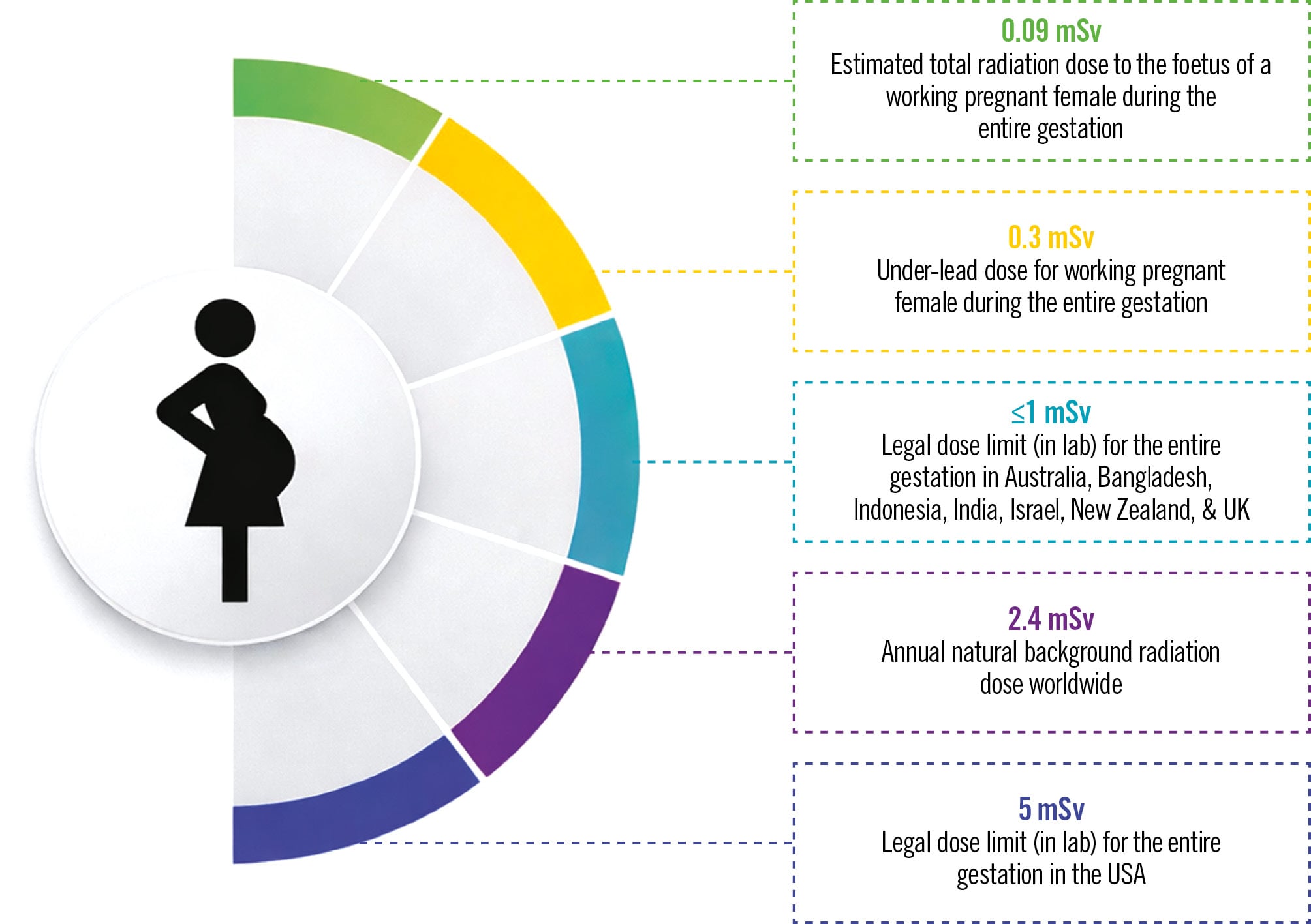
Figure 1. Foetal radiation exposure in pregnant interventionalists compared with regulatory and natural background limits. Comparison of estimated foetal radiation exposure (0.09 mSv), cumulative under-lead dose (0.3 mSv), occupational radiation dose limits during pregnancy (≤1 mSv in several countries and 5 mSv in the USA), and average annual natural background radiation (2.4 mSv). mSv: millisievert
Table 4. Radiation exposure limit for total pregnancy in different countries based on current policies.
| Radiation exposure limit for total pregnancy | Countries |
|---|---|
| ≤1 mSv | Australia, India, Bangladesh, Indonesia, Japan, United Kingdom, Israel |
| 4 mSv | Canada |
| 5 mSv | United States |
| Not allowed | Singapore, Austria, Hungary, Italy, Portugal, Romania |
| Examples of current national policies governing occupational radiation exposure during pregnancy. Limits refer to the total permitted occupational exposure during pregnancy; some countries prohibit occupational radiation exposure during pregnancy. mSv: millisievert | |
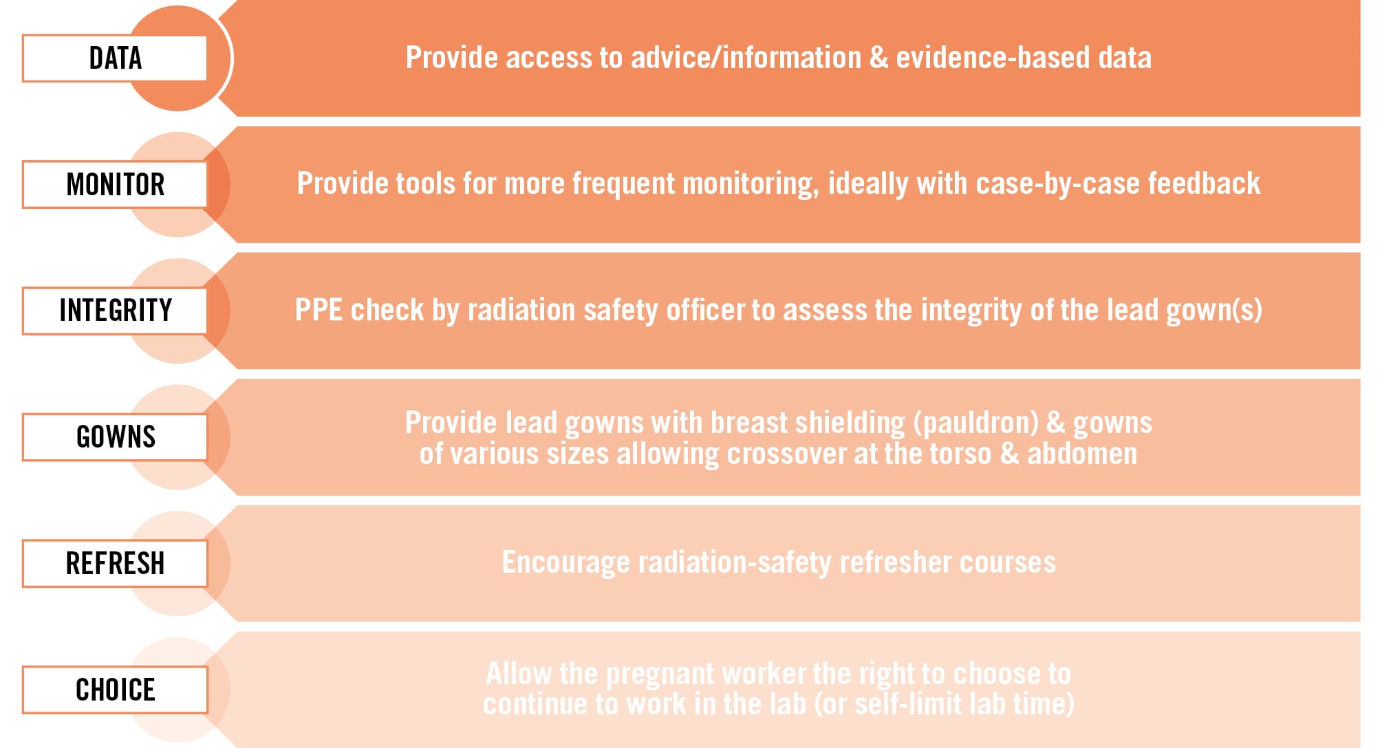
Figure 2. Strategies to enhance radiation safety for pregnant healthcare workers in the cardiac catheterisation laboratory. Key measures include access to evidence-based information, individualised radiation monitoring, regular assessment of protective equipment integrity, provision of appropriately fitted lead gowns with breast shielding, radiation safety refresher training, and informed choice regarding continued laboratory work during pregnancy. PPE: personal protective equipment
Recommendations and requirements
Operator training/education
Radiation-safety education should be mandatory for all CCL staff. Training should cover terminology, hazards, ALARA principles, practical dose-reduction techniques, and regular refreshers; structural imagers should be included.2131 CCLs should also provide routine dose reporting, preferably monthly, and formal radiation-safety programmes led by radiation protection officers.3233
Innovations in radiation protection
Novel protection systems can reduce scatter beyond traditional lead PPE. Dedicated architectural shielding is particularly important for IEs, who work close to the radiation source. Current protection systems include the following:
1. Suspended radiation protection systems: individual operator suspended radiation protection systems, such as the Zero-Gravity system, are specifically designed to enhance radiation protection to the primary operator (interventional cardiologist or electrophysiologist) while eliminating the burden of the lead apron weight.31 This system comprises a 1.0 mm suspended lead body shield that magnetically engages with a vest worn by the primary operator, attached to a 0.5 mm lead acrylic face shield. Compared with conventional lead aprons with under-table or ceiling-mounted shields, suspended radiation protection systems have been shown to provide enhanced radiation protection, with a 93.8-94.0% reduction in head-level physician radiation dose.35 Reductions in fatigue and orthopaedic injury are also noted.36 These systems are designed to protect only the first operator, and they do not decrease radiation exposure to other members of the CCL team.
2. Robotic percutaneous systems: robotic-assisted percutaneous coronary intervention (R-PCI), wherein a remote-controlled robotic system is used to undertake PCI with the operator either outside the CCL or at a remote location, has potential for addressing both a reduction in radiation exposure and the mitigation of orthopaedic issues resulting from lead apron use. R-PCI systems include the CorPath 200, the CorPath GRX system (both Corindus/Siemens Healthineers), and the R-One system (Robocath).37 With enhanced improvements, these systems resulted in significantly reduced radiation exposure to the operators (up to 96% reduction),30 with the potential to also undertake more complex lesion subsets, making them particularly useful in peripheral interventions. A randomised controlled trial38 − the PARTY trial − of a robotic-assisted system has been performed and found patient and operator radiation doses were reduced. However, the system has been withdrawn commercially.
3. Portable radiation-shielding systems: portable radiation-shielding systems, such as the RAMPART ic M1128 (Rampart), provide adjustable radiation protection shield systems, with the intention of offering “lead-free” full-bodied radiation protection for CCL staff (where local radiation policy permits).31 Preliminary data suggest that portable radiation-shielding systems may significantly reduce total body radiation exposure compared with traditional lead aprons and shields (LAS), but further independent large-scale research is required. Current early studies suggest reduced radiation exposure to body areas not traditionally protected by LAS, such as the head, and radiation attenuation to all members of the procedural team.36 The potential benefits of lead apron-free work are promising, suggesting reduced pain and fatigue, and the potential to reduce orthopaedic injury. Data showed significant protection for the primary operator (for all dosimeter locations) as well as for the second operator and scrub nurses, with reductions between 50% and 100%.32 One of the limitations of the RAMPART system is that it is not designed to attenuate radiation to the left side of the CCL, thus its utility is limited for staff who work exclusively on the left side of the patient, namely, IEs and anaesthesiologists.363738 Disposable radiation protection shields like RADPAD (Worldwide Innovations & Technologies) and SorbX (AngioSystems) are other cost-effective alternatives that can be considered to attenuate scatter radiation. They have the added advantage of being absorbent and easy to use.
4. Mobile radiation protection cabins: mobile radiation protection cabins, such as the Cathpax AIR (Lemer Pax), provide shielding of 1.0-2.0 mm of lead equivalence.39 A pilot unrandomised early feasibility study reported first-operator relative radiation exposure reduction of 78% at the chest and 70% at the wrist.39 The effect on the non-physician CCL team has not been examined.
Other commercially available radiation protection systems
Other systems include Protego (Image Diagnostics), a fixed barrier designed for lead apron-free work. A pilot cohort reported an approximately 99% reduction in operator head- and waist-level doses.40 Evidence for several proprietary systems remains largely preliminary or industry-sponsored.
Although novel systems can markedly reduce operator dose, the cost and infrastructure requirements limit their applicability across APAC, particularly in LMICs. Resource-limited CCLs should prioritise lower-cost measures such as radiation-absorbent drapes, optimised conventional shielding, and rigorous training and positioning, reserving proprietary systems for settings where resources permit. Regional procurement initiatives may improve affordability.
Room layout, protective initiatives, and the imaging echocardiographer
Ceiling-mounted shields with retractable flexible protection can accommodate C-arm rotation and improve bedside protection for imaging and interventional operators.
Structural interventions increasingly require IEs positioned close to the patient and radiation source. Transoesophageal echocardiography (TOE) provides essential soft-tissue, flow, and three-dimensional imaging, but frequent probe manipulation increases exposure, while formal radiation-safety training remains limited.41
McNamara et al reported higher per-case exposure for IEs than interventionalists despite a dedicated shield.42 Hands and feet may receive substantial exposure during probe manipulation. An additional ceiling-suspended lead shield reduced IE exposure by 82% in the study by Crowhurst et al.14
IE exposure also depends on procedure complexity, fluoroscopy time, and projection. Steep right anterior oblique (RAO) views produce high exposure,43 whereas left anterior oblique views are lower.44 Repositioning during steep RAO projections is therefore advisable. TOE guidance has been associated with an approximately threefold higher radiation dose than transthoracic echocardiography (TTE) guidance;44 fluoroscopy should also be minimised during TTE scanning.
Anaesthesiologists remain near the patient’s head during structural procedures and should be protected by mobile shielding. Although their effective dose may be lower than the interventionalist’s, appropriate shielding remains essential.
Radioprotection remains based on time, distance, and shielding. Minimising fluoroscopy, maximising distance, and using echocardiography when appropriate reduce team exposure; longer or remotely controlled ultrasound probes may offer further benefit.
New structural CCLs should incorporate dedicated IE shielding (Table 5, Table 6, Table 7), including table-mounted lower-body skirts and ceiling-mounted or upright mobile shields (Figure 3A). IEs should use appropriate eye, thyroid, and hand/forearm protection, as well as body and extremity dosimetry with regular dose review.
In practice, IE protection requires optimised positioning, advance warning of steep RAO/cranial views, consistent use of ceiling/mobile shields, appropriate PPE, body and extremity dosimetry, regular dose review, and TTE-first guidance when clinically appropriate (Table 5).
Table 5. APSIC recommendations for reducing radiation and ergonomic occupational risk in CCLs.
| Level/domain | Recommendation |
|---|---|
| Operator level – radiation hazard prevention | Educational programmes should start during training years, when habits develop, and continue throughout one’s career. |
| Adopt safe practices: limit cine runs, wear appropriate radiation protection gear, wear monitoring devices, place protective shields, and create distance from the scatter source (Table 6). | |
| Improving awareness of radiation dangers and safe practices through continuous education is a collective responsibility of individual operators, CCL personnel, hospital administration, and societies. | |
| Operator level – ergonomic/musculoskeletal hazard prevention | Use weight-distributing, two-piece pelvic-support apron designs rather than single-piece wraparound aprons. |
| Participate in structured core-strengthening/stretching exercise programmes; only 32% of WIN-APSIC respondents currently do so. | |
| Take scheduled breaks and rotate operators during long or high-volume procedure days; seek physiotherapy/occupational health review if symptomatic. | |
| Interventional echocardiographer checklist | Position self on the image-detector side near the patient’s head whenever possible; request advance notice of steep RAO/cranial views. |
| Use ceiling-suspended and mobile upright shields consistently; wear thyroid shield, glasses, leaded gloves, and forearm shield. | |
| Wear ≥2 body dosimeters plus a finger/extremity dosimeter for every case; review dose data quarterly; favour TTE-first strategies where appropriate. | |
| Institutional level | Update systems and provide protective gear, with regular checks for cracks, particularly at the seams, as a fundamental institutional requirement. |
| Continuous monitoring and regular reporting of exposure to all CCL personnel and operators (including pregnant females) is key to improving individual habits. | |
| Room layout for novel procedures is important to ensure radiation protection is optimal for all team members, including IEs and the anaesthesia team. | |
| Provide access to lightweight/composite apron materials and, where feasible, use suspended or robotic systems that remove the apron burden entirely. | |
| Consider real-time dosimetry and AI-assisted dose-optimisation/advanced collimation technology at equipment procurement or upgrade, balanced against cost and resource availability (Table 7). | |
| Policy level | Societies & authorities should elaborate a practical guide for interventional cardiologists, imagers, anaesthesiologists, and support staff to use during complex interventions: |
| Step 1 – mandate the use of dedicated shields (under-table-mounted shield, ceiling-mounted shield, mobile upright or “perforated” shield, examples shown in Figure 3) for the IEs or anaesthesiologists. | |
| Step 2 – utilise personal wearable protective devices and extremity shields (gloves, forearm/lower-limb shield) where Step 1 protection is inadequate. | |
| Step 3 – position IEs & anaesthesiologists at the head or right side of the patient whenever possible. | |
| Step 4 – limit TOE probe or endotracheal tube manipulation or transthoracic scanning during cinefluorography. | |
| Authorities should provide a central monitoring system that reports exposure doses back to institutions and individuals. | |
| Authorities/societies could incentivise institutions with safer practices. | |
| Authorities should require licensure or certification to use fluoroscopy equipment. | |
| Authorities/societies should formally recognise both radiation-related and ergonomic/musculoskeletal conditions as occupational hazards, given that awareness of this recognition is currently low across the region. | |
| APSIC recommendations for reducing radiation-related and ergonomic/musculoskeletal occupational risk at operator, interventional echocardiographer, institutional, and policy levels. AI: artificial intelligence; APSIC: Asian Pacific Society of Interventional Cardiology; CCL: cardiac catheterisation laboratory; IE: interventional echocardiographer; RAO: right anterior oblique; TOE: transoesophageal echocardiography; TTE: transthoracic echocardiography; WIN-APSIC: Women-in-Asian Pacific Society of Interventional Cardiology | |
Table 6. Practical ALARA/ALADA checklist for daily practice – containing the primary beam and scatter radiation.
| Category | Measure | Practical action in daily CCL practice |
|---|---|---|
| Primary beam/patient equipment | Collimation | Collimate tightly to the anatomical region of interest for every acquisition |
| Frame/pulse rate | Use the lowest acceptable fluoroscopy frame rate (e.g. 7.5-15 fps rather than 30 fps) as default | |
| Magnification & angulation | Avoid unnecessary magnification; minimise steep RAO/cranial views where an alternative view suffices | |
| Filtration & source distance | Maximise tube-to-patient distance; keep detector as close to the patient as possible | |
| Cine/acquisition | Restrict cine runs to essential acquisitions; use last image hold instead of repeat fluoroscopy | |
| Scatter/staff protection | Positioning | Stand on the image detector side; maximise distance from the source (inverse square law) |
| Structural shielding | Use ceiling-suspended shields, under-table lead skirts, and mobile barriers for all team members | |
| Personnel | Minimise the number of staff within the primary procedural field | |
| Personal protective equipment | Wear lead apron, thyroid shield, radiation glasses; wear extremity/finger dosimeter for high-exposure roles | |
| Monitoring | Dosimetry | Wear assigned dosimeter(s) for every case; review individual dose data at least quarterly |
| Practical daily measures to reduce the primary X-ray beam, patient exposure, and scatter radiation to catheterisation laboratory staff, consistent with ALARA/ALADA principles. ALADA: As Low As Diagnostically Acceptable; ALARA: As Low As Reasonably Achievable; CCL: cardiac catheterisation laboratory; fps: frames per second; RAO: right anterior oblique | ||
Table 7. Comparison of novel radiation protection systems, including feasibility in lower-resource (LMIC) Asia-Pacific settings.
| System | Pros | Cons | Limitations | Cost issues | LMIC feasibility |
|---|---|---|---|---|---|
| Zero-Gravity suspended radiation protection systema | ~94% reduction in head-level operator radiation; removes weight burden; reduces fatigue/orthopaedic injury; allows mobility | Only protects the primary operator; does not protect other staff | Requires compatibility with room layout and ceiling mounting | High initial installation cost; infrastructure modifications may be required | |
| Robotic PCI systems | Up to ~96% reduction in operator radiation exposure; operator can work outside radiation field; reduces orthopaedic strain; improves procedural precision | Learning curve; longer setup time; limited tactile feedback | Currently limited availability; not suitable for all lesion types; requires trained staff | Very high capital cost (purchase, maintenance, disposables) | |
| RAMPART ic M1128 portable radiation-shielding systemb | Lead-free full-body protection; protects entire procedural team; reduces exposure to areas not covered by aprons; portable and adjustable | May interfere with procedural workflow; requires space/positioning adjustments | Does not shield left side of cath lab effectively; limited protection for imagers/anaesthesiologists on the left side | Moderate-high cost; maintenance and space considerations | |
| Protego radiation protection systemc | Up to 99% reduction in operator radiation exposure; eliminates need for heavy lead aprons; comprehensive shielding barrier | Non-portable; requires fixed installation | Limited evidence, mostly single-centre; may not fit all lab layouts | High installation cost and infrastructure modification requirements | |
| Disposable drapes (RADPADd, SorbXe) & basic shielding upgrades | Low cost; no infrastructure change; immediately deployable; absorbent, easy to use | Attenuation less than dedicated barrier systems | Single-use/consumable costs accrue over time | Low | High |
| Comparison of selected contemporary radiation-protection technologies according to their potential benefits, disadvantages, practical limitations, cost considerations, and applicability in lower-resource settings. aBy TIDI Products; bby Rampart; cby Image Diagnostics; dby Worldwide Innovations & Technologies; eby AngioSystems. CCL: cardiac catheterisation laboratory; LMIC: low- and middle-income countries; PCI: percutaneous coronary intervention | |||||
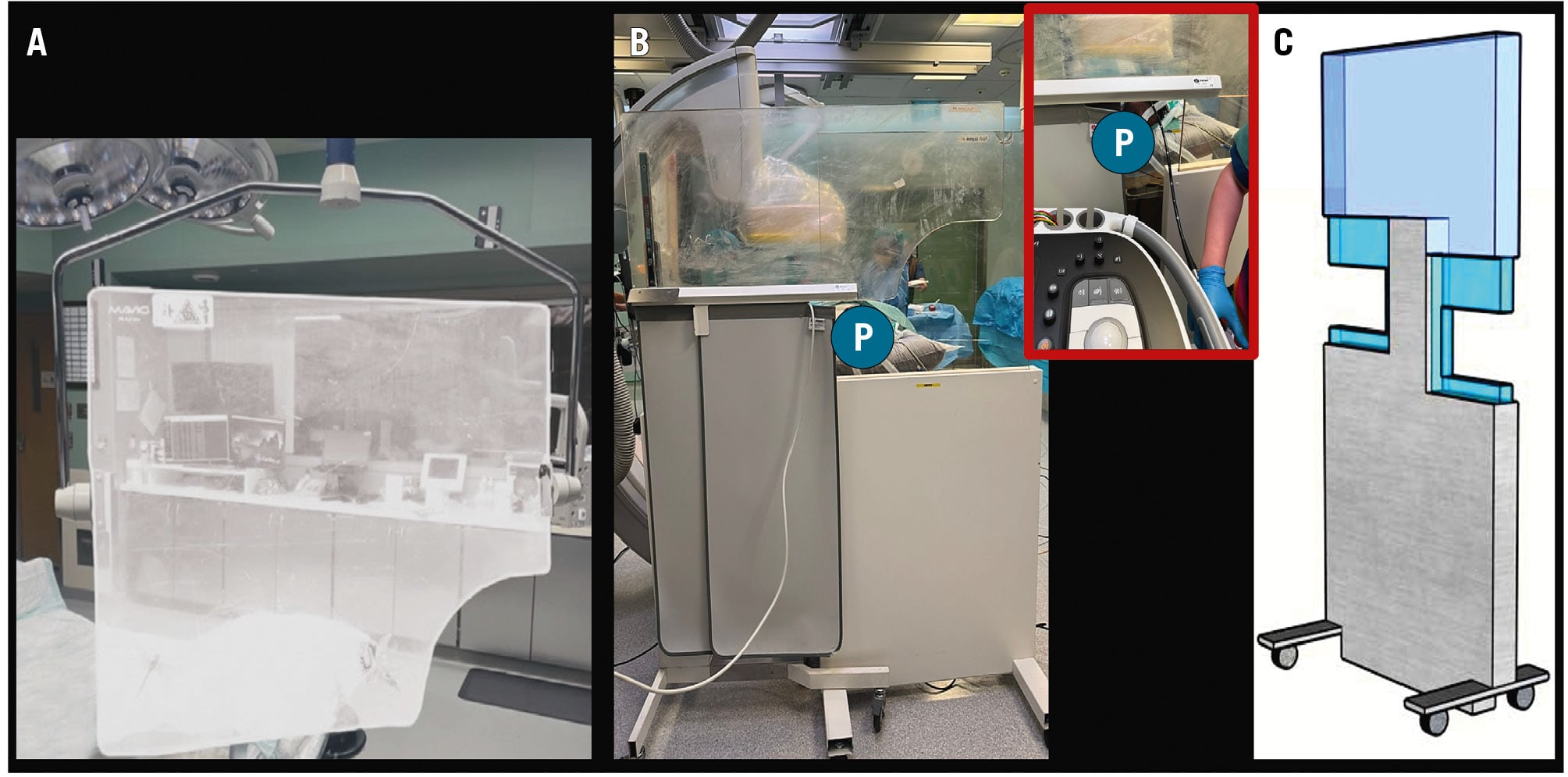
Figure 3. Examples of radiation protection shielding for interventional echocardiographers during transcatheter structural heart interventions. A) Ceiling-suspended transparent radiation shield. B) Mobile radiation protection barrier with transparent upper shielding and lead-lined lower panel, illustrating the protected position (P) behind the barrier. C) Mobile radiation protection shield with adjustable transparent upper panels and a lead-lined lower section.
Conclusions
Global and WIN-APSIC data demonstrate persistent APAC gaps in training, shielding, dosimetry, policy, and ergonomic protection. The priorities are mandatory training, standardised dose reporting, equitable access to protection, and pregnancy-inclusive policies. Emerging dose-reduction technologies may add benefit but should be implemented according to regional resources. These measures can improve safety and equity across APAC.
Conflict of interest statement
The authors have no conflicts of interest to declare.