A 66-year-old male with a history of hypertension and dyslipidaemia was admitted with ST-segment elevation myocardial infarction (STEMI). On admission, electrocardiography revealed ST-segment elevation in leads I, aVL, and V2-V6, consistent with an extensive anterior STEMI. Urgent coronary angiography demonstrated total occlusion of the proximal left anterior descending artery (Figure 1A). Primary percutaneous coronary intervention was successfully performed using an everolimus-eluting stent (SYNERGY XD, 3.5×20 mm [Boston Scientific]), restoring Thrombolysis in Myocardial Infarction grade 3 flow (Figure 1B). The door-to-balloon and onset-to-balloon times were 73 minutes and 149 minutes, respectively. Peak creatine kinase and creatine kinase-myocardial band levels reached 4,711 U/L and 442 ng/mL, respectively. Guideline-directed medical therapy was initiated, including carvedilol (2.5 mg/day), enalapril (2.5 mg/day), rosuvastatin (20 mg/day), aspirin (100 mg/day), and prasugrel (3.75 mg/day).
On hospital day 6, during cardiac rehabilitation, the patient developed palpitations and dizziness, followed by sudden loss of consciousness and cardiac arrest. Cardiopulmonary resuscitation was promptly initiated, and venoarterial extracorporeal membrane oxygenation (VA-ECMO) was established in the catheterisation laboratory. Transthoracic echocardiography revealed a pericardial effusion, prompting emergent pericardiocentesis and drainage. Repeat coronary angiography showed no evidence of in-stent thrombosis. Left ventriculography demonstrated contrast extravasation from the left ventricle (LV), indicating a blowout-type left ventricular free wall rupture (LVFWR) (Figure 1C, Moving image 1).
Due to cardiogenic shock, an Impella CP catheter (Abiomed) was inserted. The interval between cardiac arrest and initiation of VA-ECMO was 42 minutes, and Impella CP support was established 81 minutes after the arrest. The patient subsequently underwent emergency surgical repair. A median sternotomy revealed marked myocardial discolouration of the anterolateral LV wall. A myocardial laceration was identified in the territory of the first diagonal branch. Intraoperative observations of the Impella CP, in both activated and deactivated states, are shown in Figure 1D and Figure 1E. Deactivation of the Impella CP resulted in massive pulsatile bleeding from the rupture site, whereas reactivation promptly suppressed the bleeding (Moving image 2, Moving image 3). The laceration was successfully repaired with linear closure using felt strips under continued Impella CP support. Left ventricular unloading appeared to facilitate surgical repair by reducing active bleeding, thereby enabling completion of the surgery without intentional cardiac arrest.
On postoperative day 14, the Impella CP was upgraded to an Impella 5.5 (Abiomed) for long-term support. The patient was successfully weaned from VA-ECMO on postoperative day 22 and from Impella 5.5 on postoperative day 28. The patient continued rehabilitation and was transferred to another facility for prolonged rehabilitation on postoperative day 48.
Blowout-type LVFWR is a catastrophic and life-threatening complication of acute myocardial infarction, associated with extremely high mortality rates.1 Although mechanical circulatory support with intra-aortic balloon pumping, VA-ECMO, or a combination of the two has previously been the conventional treatment approach in post-infarction mechanical complications, an increasing number of reports have recently suggested that the use of Impella may be effective in patients with post-infarction mechanical complications as a bridge to surgical repair.23 The Impella system provides continuous LV unloading, which may help to reduce myocardial wall stress and reduce LV intracavitary pressure, potentially preventing rerupture at the repair site.4567 The bleeding volume from the rupture site is determined by the pressure gradient between the LV cavity and pericardial space. We hypothesise that LV unloading reduced bleeding by decreasing the pressure gradient through reduction of the LV intracavitary pressure. Although the use of the Impella system in mechanical complications of acute myocardial infarction has been previously reported,7 this is the first case to visually demonstrate real-time LV unloading by Impella during surgical repair.
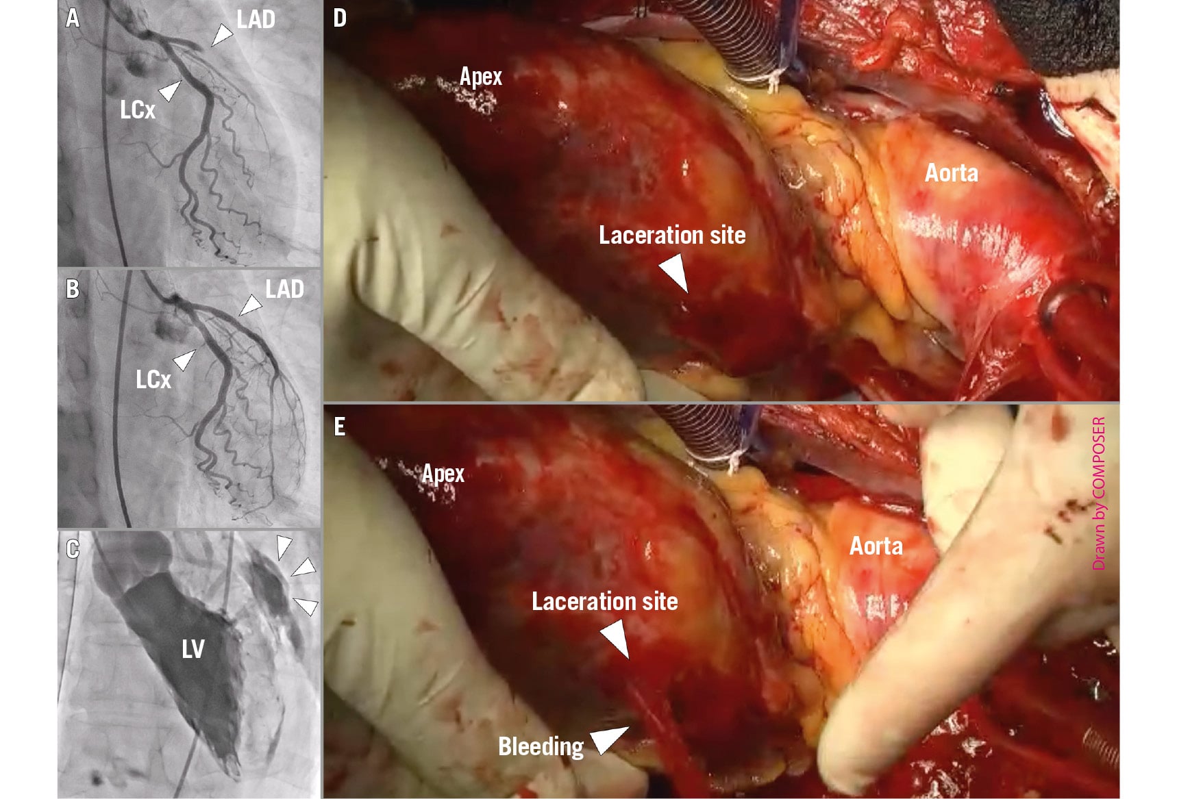
Figure 1. Urgent percutaneous coronary intervention and intraoperative observations demonstrating left ventricular pressure unloading by Impella CP support. A) Coronary angiography demonstrated total occlusion of the proximal left anterior descending artery. B) Following implantation of an everolimus-eluting stent (SYNERGY XD, 3.5×20 mm), final angiography showed TIMI grade 3 flow. C) On hospital day 6, following cardiac arrest during rehabilitation and establishment of VA-ECMO, left ventriculography revealed a blowout-type LVFWR, as indicated by the white arrowheads. D) After a median sternotomy, no active pulsatile bleeding was observed under Impella CP support. E) Temporary deactivation of the Impella CP device resulted in active pulsatile bleeding from the laceration site. LAD: left anterior descending artery; LCx: left circumflex artery; LV: left ventricle; LVFWR: left ventricular free wall rupture; TIMI: Thrombolysis in Myocardial Infarction; VA-ECMO: venoarterial extracorporeal membrane oxygenation
Conflict of interest statement
The authors have no conflicts of interest to declare.