We report two cases of percutaneous device closure of ascending aortic pseudoaneurysms. First, we present a 31-year-old male with Marfan syndrome and multiple aortic dissections who required open and endovascular aneurysm graft repair. He underwent a Bentall procedure with mechanical aortic valve replacement and coronary reimplantation. A computed tomography (CT) aortogram post-Bentall surgery showed an ascending aorta pseudoaneurysm anterior to the conduit graft measuring 60×40 mm, with a pseudoaneurysm neck measuring 8 mm. Percutaneous device closure was chosen because of the high open surgical risk. A 6 Fr 120 cm Judkins right 4 (JR4) catheter was carefully manipulated up to the ascending aorta using a GLIDEWIRE (Terumo) (Moving image 1). Thereafter, a JR4 catheter was advanced into the pseudoaneurysm using GLIDEWIRE support. This was exchanged for a stiff support wire, followed by railroading of a 7 Fr 90 cm Pinnacle Destination delivery sheath (Terumo) into the pseudoaneurysm (Moving image 2), where an Amplatzer Septal Occluder (ASO; Abbott) 10 mm device was then placed. An ascending aortogram showed successful occlusion of the pseudoaneurysm, which was confirmed on a CT aortogram 3 months later (Figure 1A – Figure 1B – Figure 1C – Figure 1D – Figure 1E).
Second, we present the case of a 60-year-old female with a history of aortic coarctation repair. A CT aortogram showed a 53×43 mm pseudoaneurysm arising from the inferior aspect of the aortic isthmus. The pseudoaneurysm neck measured 10-12 mm. Open pseudoaneurysm repair was abandoned because of aortic calcifications. Percutaneous pseudoaneurysm closure was performed via right femoral access. A 5 Fr Cobra catheter was positioned at the pseudoaneurysm neck using a GLIDEWIRE. However, there was insufficient support to advance the 8 Fr Amplatzer delivery catheter (Abbott). The GLIDEWIRE was looped out of the pseudoaneurysm and into the descending aorta for support (Moving image 3). Subsequently, a 125 cm 4 Fr multipurpose diagnostic catheter was advanced through the GLIDEWIRE and delivery sheath into the pseudoaneurysm (Moving image 4). With this mother-in-child technique, there was sufficient support to advance the delivery sheath. A 16 mm Amplatzer vascular plug II (AVP II; Abbott) was deployed (Moving image 5). The final aortogram showed good closure, and a CT aortogram 6 months later confirmed pseudoaneurysm occlusion (Figure 1F – Figure 1I – Figure 1J). Both patients remain stable with no pseudoaneurysm recurrence at 2-year follow-up.
Aortic pseudoaneurysm carries a high mortality rate of up to 61%.1 In patients with high surgical risk, percutaneous pseudoaneurysm closure may be lifesaving, with procedural success rates of 90-100%.2 Nevertheless, careful consideration must be emphasised3 as procedural complications such as perforation, dissection, and stroke can occur.4 If the pseudoaneurysm cavity is small, there may be insufficient wire support to advance the large-bore delivery catheter. Techniques showcased in these cases include looping the wire within the cavity or advancing a smaller, longer catheter as a railroad. The choice of closure device depends on the size of the pseudoaneurysm and its neck. We chose the 10 mm ASO device for the first case, as the aneurysm cavity was large with a narrow neck (8 mm). For the second, the 16 mm AVP II was chosen for its smaller discs, which were better suited for the small aneurysm cavity with a wide neck (12 mm). An individualised approach for preprocedural planning is key to anticipating potential challenges and aiding in device selection and sizing.
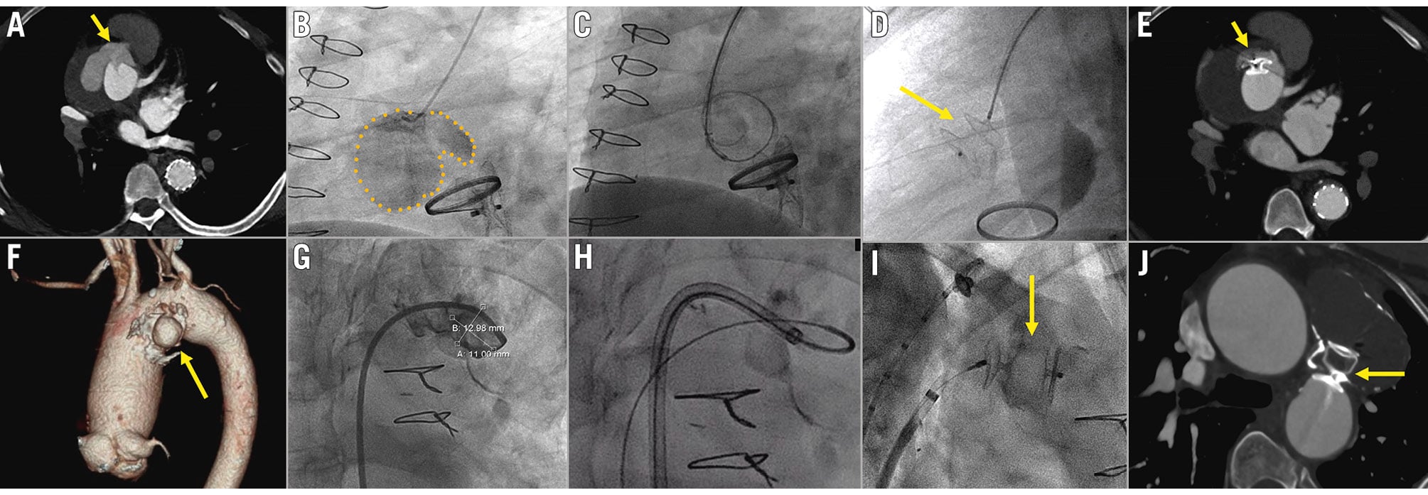
Figure 1. Two cases of pseudoaneurysm closure. Patient 1: A) CT aortogram 10 months post-Bentall surgery showing an interval development of an aortic root pseudoaneurysm arising just superior to the mechanical aortic valve and anterior to the conduit graft. A defect is seen at the expected location of the previously reimplanted SVG-RCA graft, indicating the possible site of communication between the graft and pseudoaneurysm (yellow arrow). B) Angiography showed a large pseudoaneurysm near the ostium of the SVG-RCA conduit (dotted yellow line). C) Fluoroscopy image showing that the pseudoaneurysm was cannulated with the JR4 catheter and the pigtail wire introduced. The 90 cm delivery sheath was then railroaded into the pseudoaneurysm. D) Fluoroscopy image showing the deployment of the Amplatzer Septal Occluder (ASO; yellow arrow) within the pseudoaneurysm. E) CT aortogram 3 months post-procedure showed successful occlusion of the pseudoaneurysm arising from the previous SVG-RCA reimplantation site. Yellow arrow pointing to the ASO. Patient 2: F) CT aortogram with three-dimensional reconstruction showing the pseudoaneurysm (yellow arrow) arising from the inferior aspect of the aortic isthmus. G) Aortogram of the calcified pseudoaneurysm with depth of the cavity measuring 13 mm and width of 11 mm. H) Fluoroscopy image showing the Amplatzer delivery catheter advancing over the multipurpose catheter and GLIDEWIRE, both looped inside the pseudoaneurysm. I) Fluoroscopy image showing deployment of the Amplatzer vascular plug (AVP; yellow arrow) in the mouth of the pseudoaneurysm orifice. J) Interval CT aortogram 6 months post-device closure showing the AVP (yellow arrow) in the neck of the pseudoaneurysm. No leakage of contrast material is seen into the pseudoaneurysm, indicating successful closure. The aneurysm sac is completely thrombosed. CT: computed tomography; JR: Judkins right; RCA: right coronary artery; SVG: saphenous vein graft
Conflict of interest statement
J. Yap has received speaker honoraria from Abbott, Biosensors, Boston Scientific, Edwards Lifesciences, Medtronic, and Johnson & Johnson. The other authors have no conflicts of interest to declare.