Percutaneous coronary intervention (PCI) is becoming more complex, given the increasingly advanced age of the population and concomitant increases in morbidity indices and both atherosclerotic and non-atherosclerotic risk factors.123 Complex PCI is typically associated with low rates of procedural success and high rates of recurrence or major adverse cardiac events.45 A careful diagnostic workup of the patient is key to successful complex PCI. In this context, the routine implementation of intravascular imaging (IVI) in complex coronary artery disease is a crucial part of preinterventional lesion assessment and postinterventional stent optimisation.
Recently, the Randomized Controlled Trial of Intravascular Imaging Guidance Versus Angiography-Guidance on Clinical Outcomes After Complex Percutaneous Coronary Intervention (RENOVATE-COMPLEX-PCI) showed that, among patients with complex coronary artery lesions, IVI-guided PCI led to a lower risk of a composite of death from cardiac causes, target vessel-related myocardial infarction (MI), or clinically driven target vessel revascularisation (TVR) than angiography-guided PCI.6 However, patients with complex lesion anatomy undergoing coronary stenting may experience different risks and outcomes according to procedural complexity. Given this context, patients undergoing complex PCI procedures may derive greater benefits from IVI guidance compared with those undergoing less complex PCI. In this study, we evaluated the effectiveness of IVI-guided versus angiography-guided complex PCI in improving clinical outcomes according to PCI procedural complexity.
Methods
Study design
This study was a post hoc analysis of the RENOVATE-COMPLEX-PCI (ClinicalTrials.gov: NCT03381872), an investigator-initiated, randomised, open-label, multicentre, superiority trial conducted at 20 sites in the Republic of Korea. Briefly, patients aged ≥19 years who underwent PCI for complex coronary artery lesions were enrolled. Complex coronary artery lesions were defined as true bifurcation lesions with a side branch ≥2.5 mm in size, chronic total occlusions (CTOs), unprotected left main artery disease, long coronary lesions, multivessel PCI requiring multiple stents, in-stent restenosis, severe calcified lesions, or coronary ostial lesions. The key exclusion criteria were cardiogenic shock at presentation and known hypersensitivity to or contraindications for the use of aspirin, clopidogrel, prasugrel, ticagrelor, heparin, everolimus, or contrast media.6
This study was conducted in accordance with the principles of the Declaration of Helsinki. The institutional review board approved the trial protocol of each participating site. The authors confirm that patient consent forms were obtained for this article.
Randomisation and treatment
Patients with treatable complex coronary artery lesions were randomised in a 2:1 ratio to undergo IVI-guided or angiography-guided PCI. A standard PCI technique was used, and the choice of imaging modality and timing of its use were left to the discretion of the operators. Although IVI can be used at any stage of the PCI procedure (before, during, or after PCI), post-PCI imaging is required to ensure optimal stent deployment and adjustment of the treated segment. Protocols for IVI acquisition and procedural optimisation were established based on the latest expert consensus recommendations.7 After the index procedure, dual antiplatelet therapy was advised for a duration of at least 3-6 months in patients with stable ischaemic heart disease and for 6-12 months in patients with acute coronary syndrome (ACS). However, the choice of loading dose, maintenance dose, and overall duration of dual antiplatelet therapy was left to the discretion of the treating physician. Irrespective of the group assignment, the medical treatment of each patient followed the most up-to-date clinical guidelines.89
Endpoints and definitions
The primary endpoint was target vessel failure (TVF), defined as a composite of cardiac death, target vessel-related MI, or clinically driven TVR. The secondary endpoints included the individual components of the primary endpoint: cardiac death, target vessel-related MI, and TVR. Spontaneous MI was defined based on the Third Universal Definition,10 whereas procedure-related MI was defined based on the guidelines set by the Society for Cardiovascular Angiography and Interventions.11 Deaths from unknown causes were categorised as cardiac deaths, with other endpoints defined in accordance with the Academic Research Consortium criteria.12
Stratification of procedural complexity
In the current substudy, all patients enrolled in the RENOVATE-COMPLEX-PCI trial were stratified into three groups of low, moderate, and high risk, according to PCI procedural complexity (Supplementary Figure 1) as assessed by the criteria of the 2023 European Society of Cardiology (ESC) guidelines for ACS management (Supplementary Figure 2).13 The high-risk group consisted of patients who met at least one of the clinical risk criteria (diabetes mellitus; chronic kidney disease [CKD], which was defined as a glomerular filtration rate between 15 mL/min/1.73 m² and 60 mL/min/1.73 m²; prior MI; peripheral artery disease [PAD]; age <45 years; or multivessel coronary artery disease) and at least one of the angiographic risk criteria (≥3 stents implanted, ≥3 lesions treated, total stent length >60 mm, left main artery PCI, CTO, or bifurcation lesions treated with 2 stents). Patients in the moderate-risk group met at least one of the following clinical risk criteria: diabetes mellitus, CKD, prior MI, or PAD. The low-risk group included patients who did not fulfil the criteria for the high- or moderate-risk groups.
Statistical analysis
All analyses were performed on an intention-to-treat basis. Categorical variables are reported as numbers and relative frequencies (%) and were compared using the χ2 and Fisher’s exact tests. Continuous variables are reported as mean±standard deviation and were compared using the Student’s t-test or Mann-Whitney U test. Kaplan-Meier analyses were performed for time-to-event outcomes, with intervention effects estimated using Cox proportional hazard regression models; the results are presented as hazard ratios (HRs) with 95% confidence intervals (CIs). Covariates that were either statistically significant in the univariate analysis (p<0.10) or considered clinically important were included in the multivariate models. All probability values were two-sided, and p<0.05 was considered statistically significant. Statistical analyses were performed using SPSS Statistics software, version 20.0 (IBM).
Results
Baseline characteristics
The study population consisted of 1,639 patients, of whom 370 (22.6%), 232 (14.2%), and 1,037 (63.3%) were classified into the low-, moderate-, and high-risk groups, respectively (Supplementary Figure 1). The baseline clinical and procedural characteristics according to PCI complexity are shown in Supplementary Table 1. Some clinical and angiographic characteristics differed significantly across the three risk groups. The patients in the moderate- and high-risk groups were older, whereas hypertension and diabetes were more common in the high-risk group than in the other groups. CKD was more common in the moderate- and high-risk groups than in the low-risk group. The patients in the moderate- and high-risk groups more frequently had multivessel disease, left main artery involvement as the culprit lesion, true bifurcation lesions, and CTOs. Procedural metrics, including total stent length, total contrast volume, and procedural time, were significantly greater in the high-risk group than in the other groups. Procedural success rates were high in all groups but were slightly lower in the high-risk group than in the other groups. Medication use, including clopidogrel, prasugrel, and ticagrelor, varied significantly across the groups, with higher use of beta blockers and renin-angiotensin system inhibitors in the moderate- and high-risk groups than in the low-risk group. Finally, the duration of hospitalisation was significantly longer in the high-risk group than in the other groups.
Comparison of clinical outcomes based on procedural complexity
At a median follow-up of 2.1 years (interquartile range 1.4-3.0 years), the primary endpoint of cardiac death, target vessel-related MI, or clinically driven TVR occurred in 18 patients (4.9%) in the low-risk group, 19 (8.2%) in the moderate-risk group, and 99 (9.5%) in the high-risk group. The risk of the primary endpoint increased stepwise in the moderate-risk (adjusted HR 1.369, 95% CI: 0.705-2.660) and high-risk groups (adjusted HR 1.619, 95% CI: 0.963-2.723) compared with that in the low-risk group (Figure 1).
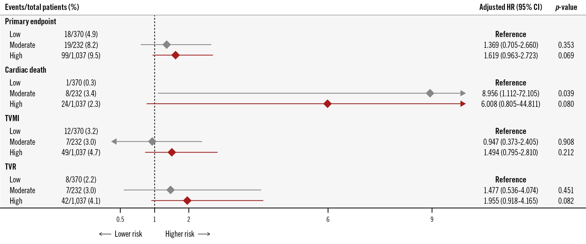
Figure 1. Adjusted risk of clinical outcomes by procedural complexity. The primary endpoint, TVF, was defined as a composite of cardiac death, TV-related MI, or clinically driven TVR. Event rates and adjusted HRs for TVF and its individual components are presented across the low-, moderate-, and high-risk groups, classified according to PCI procedural complexity. A stepwise increase in the adjusted risk of TVF was observed in the moderate-risk (HR 1.369, 95% CI: 0.705-2.660) and high-risk groups (HR 1.619, 95% CI: 0.963-2.723) compared with that in the low-risk group. CI: confidence interval; HR: hazard ratio; MI: myocardial infarction; PCI: percutaneous coronary intervention; TV: target vessel; TVMI: target vessel-related myocardial infarction; TVF: target vessel failure; TVR: target vessel revascularisation
Comparison of clinical outcomes in IVI-guided PCI versus angiography-guided PCI by procedural complexity
In the angiography-guided PCI group, TVF occurred in 5.6%, 13.1%, and 12.4% of the patients in the low-, moderate-, and high-risk groups, respectively. In the IVI-guided PCI group, the rates of TVF were 4.5%, 5.4%, and 8.2% of the patients in the low-, moderate-, and high-risk groups, respectively. The unadjusted HRs for TVF showed a significant risk reduction in the moderate-risk (HR 0.389, 95% CI: 0.157-0.968; p=0.042) and high-risk groups (HR 0.645, 95% CI: 0.433-0.960; p=0.031) with IVI-guided PCI (Table 1). After adjustment, the HR remained significant in the moderate-risk (adjusted HR 0.374, 95% CI: 0.150-0.935; p=0.035) and high-risk groups (adjusted HR 0.633, 95% CI: 0.424-0.945; p=0.025). The effect of IVI-guided PCI varied according to PCI procedural complexity (adjusted p for interaction=0.009), showing a significant benefit of using IVI in the patients in the moderate-risk and high-risk groups compared with those in the low-risk group (Figure 2).
Table 1. Comparison of clinical outcomes between IVI-guided PCI and angiography-guided PCI by procedural complexity.
| ESC criteria/ randomisation | Low risk (n=370) | Moderate risk (n=232) | High risk (n=1,037) | Interaction p-value |
|---|---|---|---|---|
| Angiography-guided PCI | 7/125 (5.6) | 11/84 (13.1) | 42/338 (12.4) | |
| Intravascular imaging-guided PCI | 11/245 (4.5) | 8/148 (5.4) | 57/699 (8.2) | |
| Unadjusted HR for TVF (95% CI) | 0.795 (0.308-2.052) p=0.636 | 0.389 (0.157-0.968) p=0.042 | 0.645 (0.433-0.960) p=0.031 | 0.013 |
| Adjusted HR for TVF* (95% CI) | 0.735 (0.282-1.914) p=0.528 | 0.374 (0.150-0.935) p=0.035 | 0.633 (0.424-0.945) p=0.025 | 0.009 |
| Data are n/N (%) or HR (95% CI). *Adjusted for ACS, previous heart failure, ejection fraction, and randomised group (IVI-guided PCI versus angiography-guided PCI). ACS: acute coronary syndrome; CI: confidence interval; ESC: European Society of Cardiology; HR: hazard ratio; IVI: intravascular imaging; PCI: percutaneous coronary intervention; TVF: target vessel failure | ||||
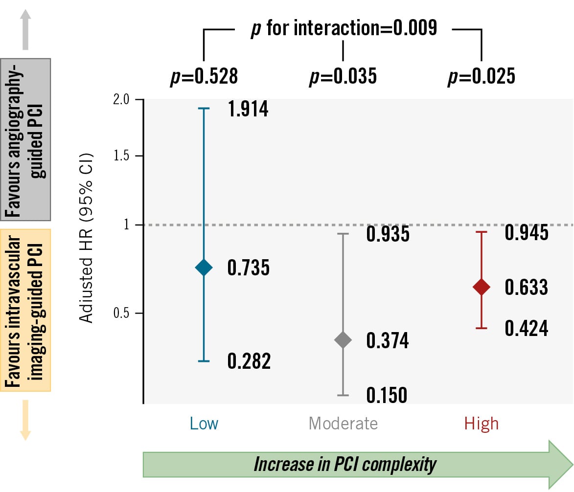
Figure 2. Adjusted risk of target vessel failure with IVI-guided versus angiography-guided PCI by procedural complexity. Adjusted hazard ratios for TVF comparing IVI-guided with angiography-guided PCI are shown across the low-, moderate-, and high-risk groups, classified according to PCI procedural complexity. The clinical benefit of IVI guidance became progressively more pronounced with increasing complexity (p for interaction=0.009), achieving statistical significance in the moderate- and high-risk groups. CI: confidence interval; HR: hazard ratio; IVI: intravascular imaging; PCI: percutaneous coronary intervention; TVF: target vessel failure
Discussion
Among patients enrolled and randomised in the RENOVATE-COMPLEX-PCI trial, we found that those undergoing more complex PCI had a higher rate of subsequent ischaemic events compared with those undergoing standard PCI. The present study also found that the benefit of IVI-guided PCI increased additively with higher PCI procedural complexity (Central illustration).
Several hypotheses may explain these findings: (1) complex PCI is associated with higher risks of death, MI, and unplanned revascularisation.45 A careful diagnostic workup of the patient is the cornerstone for successful complex PCI procedures. In this respect, the routine assessment of IVI in complex coronary artery disease is pivotal for the diagnostic workup. Ultimately, for complex PCI, IVI should be a valuable adjunctive tool for preinterventional lesion assessment.141516 2) Stenting of complex lesions may increase the risks of stent malapposition, incomplete lesion coverage, and delayed endothelialisation, all of which, in turn, are involved in the enhancement of thrombotic propensity within the target vascular segment.17 In this context, routine assessment of IVI in complex coronary artery disease is essential for postinterventional stent optimisation. Recently, Witzenbichler et al showed that the benefits of intravascular ultrasound (IVUS) were especially evident in patients with ACS and complex lesions in ADAPT-DES.18 Bavishi et al also showed in their meta-analysis that IVUS-guided PCI led to significant reductions (by 36%) in the rates of major adverse cardiac events, in comparison with angiography guidance, with maximal benefit in patients with ACS, diabetes mellitus, and long lesions.19
In addition to lesion complexity, clinical factors play an important role in PCI outcomes. Conditions such as diabetes mellitus, CKD, or a history of MI are known to accelerate the progression of coronary artery disease and complicate treatment. The interplay between these clinical comorbidities and anatomical complexity has been reported to have a significant impact on procedural success and long-term outcomes.20 Considering these points, in our study, we adopted the ESC criteria13 to quantify procedural complexity by incorporating both clinical and anatomical factors. This approach allowed us to categorise patients into low-, moderate-, and high-risk groups according to procedural complexity, thus offering a more comprehensive evaluation of the impact of IVI-guided PCI.
Major scientific cardiology societies have recommended the use of IVI to guide and optimise stent implantation in selected cases, including complex PCI. Previous consensus and guideline documents had assigned this a Class IIa recommendation.7921 However, more recently, the 2025 ACC/AHA/ACEP/NAEMSP/SCAI Guideline for the Management of Patients With Acute Coronary Syndromes has upgraded the use of intravascular imaging to a Class I, Level of Evidence A recommendation in ACS patients with complex coronary lesions.22 Despite these recommendations, the overall adoption of IVI remains small, with current estimates indicating usage in <15% of all PCI procedures in the United States.15 Various explanations have been proposed for the underuse of IVI, including unfamiliarity with imaging equipment, knowledge gaps in image interpretation, recognition that increased procedure time adversely affects workflow efficiency, and a lack of adequate financial reimbursement.23 In particular, in the setting of a complex procedure, hesitation in the use of the equipment may also be considered an important factor. Given the remaining difficulties in the widespread adoption of IVI, additional evidence to reinforce its benefits is needed. This finding may be helpful for decision-making in real-world clinical practice. Our comprehensive approach to assessing PCI complexity strengthens the conclusions of this study and provides a clearer understanding of the incremental benefits of IVI in different patient populations.
Two recent trials, OPTIMAL24 and IVUS-CHIP,25 yielded opposite results to the above findings. One of the randomised trials, OPTIMAL, showed no significant benefit of IVUS over angiography for guiding PCI in revascularisation of left main stenosis. The other, IVUS-CHIP, was a larger trial enrolling patients with a broader range of complex coronary artery lesions, and it too revealed no advantage of IVUS-guided PCI on the key endpoints. Although IVUS did not reduce the risk of events in these two trials, this conclusion should be restricted to procedures performed by expert IVUS operators. This is because IVUS experience improves the imaging skills of interventionalists, likely allowing experienced interventionalists to achieve good results with any type of imaging, whether with IVUS or angiography. However, the bottom line is that imaging is extremely valuable in developing an interventionalist’s PCI practice.
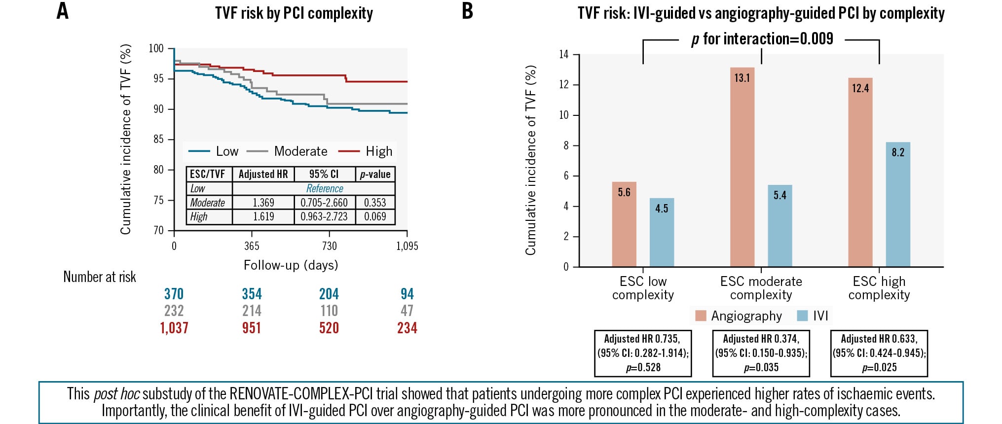
Central illustration. Clinical impact of IVI-guided PCI across procedural complexity. TVF risk by (A) PCI complexity and (B) IVI-guided vs angio-guided PCI. CI: confidence interval; ESC: European Society of Cardiology; HR: hazard ratio; IVI: intravascular imaging; PCI: percutaneous coronary intervention; RENOVATE-COMPLEX-PCI: Randomized Controlled Trial of Intravascular Imaging Guidance Versus Angiography-Guidance on Clinical Outcomes After Complex Percutaneous Coronary Intervention; TVF: target vessel failure
Limitations
This study has some limitations. First, randomisation in the RENOVATE-COMPLEX-PCI trial was not stratified by procedural complexity, as this was a post hoc analysis of a randomised clinical trial. In addition, the sample size was small, especially in the moderate- and high-complexity subgroups. Consequently, the analysis may lack sufficient statistical power to detect differences in the primary endpoint between the study groups. In the same context, in the low procedural complexity subgroup, no significant difference in TVF was observed between IVI-guided and angiography-guided PCI. The small sample size and low event rate limit the reliability of this observation. Therefore, comparisons based on procedural complexity should be regarded as hypothesis-generating. Second, procedural complexity was defined using the ESC criteria in this substudy. However, the ESC criteria were developed to identify patients with ACS at moderate or high thrombotic risk, for whom extended treatment with a second antithrombotic agent might be considered after PCI;2627 therefore, the ability of these criteria to define PCI procedural complexity has not been validated. Finally, the HRs are borderline significant with wide CIs.
Conclusions
Patients undergoing complex PCI procedures are at a substantially higher risk of ischaemic events, and this risk increases with PCI procedural complexity. Among patients who underwent complex PCI, IVI-guided PCI significantly reduced the risk of cardiac ischaemic events, compared with angiography-guided PCI, with an increased magnitude of benefit with greater procedural complexity. Our study suggests a potential benefit of using IVI in procedural high-risk PCI patients to reduce long-term adverse outcomes.
Declaration of generative AI use
The authors did not use generative artificial intelligence (AI) or AI-assisted tools during the manuscript preparation process.
Impact on daily practice
PCI complexity is associated with an increased risk of adverse cardiac events. In our study, the benefit of imagingguided PCI became more pronounced as PCI complexity increased, highlighting the potential value of risk-adapted imaging strategies.
Conflict of interest statement
J.-Y. Hahn reports receiving grants from Abbott and Boston Scientific during the conduct of the study; and grants from the National Evidence-based Healthcare Collaborating Agency, the Ministry of Health & Welfare, Republic of Korea, Biosensors, Biotronik, Daiichi Sankyo, Dong-A ST, Hanmi Pharmaceutical, and Medtronic, outside the submitted work. C.-W. Nam reports receiving grants from Abbott outside the submitted work. The other authors have no conflicts of interest to declare.