Transcatheter aortic valve implantation (TAVI) has become a preferred approach to treat symptomatic severe aortic stenosis (AS). As TAVI expands into younger and lower-risk populations, expectations increasingly emphasise durable, patient-centred outcomes, thereby reinforcing the need for reproducible implantation strategies and continued procedural refinement for ongoing safety and efficiency.
For supra-annular self-expanding TAVI (e.g., with the Evolut platform [Medtronic]), standardised fluoroscopic strategies and protocolised pathways have gained momentum, with evidence that a codified technique can improve implantation consistency and clinically meaningful outcomes, such as a reduction in permanent pacemaker implantation (PPI) rates.123 In particular, the Optimize PRO Study and the Optimize PRO Evolut FX addendum demonstrated that a standardised Evolut implantation pathway can achieve low PPI rates, indicating the clinical value of technique standardisation in contemporary self-expanding TAVI.23 However, many prospective reports in this setting emphasise global workflow measures, such as total procedure room occupancy,2 which are influenced by non-device-related steps. Accordingly, these measures may be less specific to the device-manipulation step that procedural refinement intends to improve upon. In this context, a deployment-centric time metric that focuses on device manipulation may provide a more modifiable and actionable endpoint for iterative procedural refinement and quality improvement.
In this context, we implemented a semi-rapid pacing-assisted deployment workflow for the Evolut platform to improve efficiency during the key deployment phase while preserving standard depth-assessment checkpoints (Central illustration). In this single-centre before-and-after implementation study, we evaluated its mechanistic validity focusing on device time − defined as the interval from initial advancement of the delivery system to final release of the transcatheter heart valve (THV) from the delivery system − rather than broader time metrics dominated by room logistics. We further examined whether this deployment refinement was associated with early valve-related and clinical outcomes, including short-term mortality, stroke, paravalvular leak (PVL), and PPI.
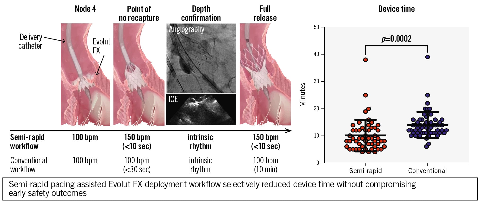
Central illustration. Impact of semi-rapid pacing on Evolut FX deployment time. The semi-rapid workflow alternates 100 bpm for checkpoint confirmation and 150 bpm for brief predefined deployment phases, reducing device time while preserving depth-confirmation steps. bpm: beats per minute; ICE: intracardiac echocardiography
Methods
Study design and oversight
Our prospectively collected, retrospectively analysed study enrolled consecutive patients with severe aortic stenosis who underwent transfemoral TAVI with the Evolut FX system (Medtronic) between March 2023 and January 2025, for whom procedural timing data were available. Written informed consent was obtained from all participants for the present registry. The study protocol complies with the Declaration of Helsinki and was approved by the Institutional Review Board of Juntendo University Hospital (IRB 17-263).
Study groups
Participants were categorised according to implantation strategy and study period. The conventional group (March 2023-March 2024) underwent conventional stepwise Evolut deployment under ventricular pacing at 100 beats per minute (bpm) throughout the deployment sequence. The semi-rapid group (April 2024-January 2025) underwent a pacing-assisted strategy alternating between 100 bpm and 150 bpm (“semi-rapid pacing method”).
Procedural details and implantation strategies
In the conventional approach, the Evolut valve was advanced and deployed in a stepwise manner under 100 bpm pacing. The device was deployed gradually to the point of no recapture, followed by confirmation of position and depth using angiography, with intracardiac echocardiography (ICE) used when available. Final valve release was then completed while maintaining 100 bpm pacing, prioritising controlled depth optimisation during deployment.
In the semi-rapid pacing method (Figure 1), pacing at 100 bpm and 150 bpm was alternated to combine controlled assessment with rapid advancement during time-sensitive steps. After the delivery system crossed the native aortic valve, the valve was initially deployed to node 4 at 100 bpm and the position was verified by contrast fluoroscopy. Node 4 denotes a prespecified intermediate stage in the stepwise Evolut FX deployment sequence, at which point partial expansion provides stable fluoroscopic visualisation for standardised position and depth checks. After attachment to the non-coronary cusp, pacing was increased to 150 bpm to rapidly advance device deployment to the point of no recapture over a few seconds. Backup pacing at 100 bpm was then applied, and implantation depth was assessed using ICE in addition to angiographic guidance with the cusp-overlap and left anterior oblique views. The temporary pacing rate was transiently reduced to confirm the patient’s intrinsic heart rate without pacing. When position or depth correction was required, recapture was performed after reassessment of implantation depth and valve position and before final valve release. After confirming an appropriate implantation depth, pacing was again increased to 150 bpm to facilitate rapid final deployment and valve release (typically within 5-10 seconds). This alternation between 100 bpm and 150 bpm was intended to preserve depth-confirmation checkpoints while shortening the interval between them, supported by multimodality intraprocedural imaging (angiography and ICE). The 150 bpm rate was selected pragmatically as an intermediate pacing rate intended to transiently reduce stroke volume and valve motion during predefined time-sensitive deployment steps while maintaining haemodynamic stability. Full rapid ventricular pacing (>180 bpm) was avoided because of concern for haemodynamic intolerance. This pacing-assisted semi-rapid deployment workflow was implemented at our institution based on a concept initially introduced in contemporary Evolut implantation practice by Tang and colleagues and was subsequently standardised and optimised within our institutional protocol.
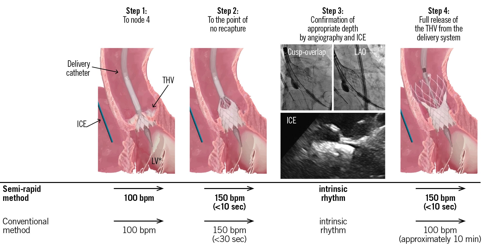
Figure 1. Semi-rapid pacing-assisted deployment workflow for Evolut FX. Schematic comparison of the stepwise implantation workflows. Step 1: advancement to node 4; step 2: advancement to the point of no recapture; step 3: confirmation of appropriate implantation depth by angiography (cusp-overlap and left anterior oblique [LAO] views) and intracardiac echocardiography (ICE), when used; step 4: full release. Recapture, when required for position or depth correction, was performed after step 3 and before step 4. In the conventional workflow, pacing at 100 bpm is maintained throughout deployment, whereas the semi-rapid method alternates 100 bpm for checkpoint confirmation and 150 bpm for brief, predefined rapid deployment phases. THV: transcatheter heart valve
Endpoints
The primary outcomes were procedure-related metrics, including operation time, procedure time, device time, fluoroscopy time, and contrast volume. Operation time was defined as the room-in to room-out interval (from patient entry into the procedure room to exit) and was extracted from the anaesthesia record. Procedure time was the access-to-haemostasis time, defined as the interval from vascular access (skin puncture) to completion of vascular haemostasis, extracted from the anaesthesia record. Device time was defined as the duration from initial advancement of the delivery system to final release of the THV from the delivery system, as captured by the angiography system timestamps. Fluoroscopy time was obtained from the angiography system. Contrast volume represented the total contrast administered during the procedure and was obtained from angiography system logs and/or procedural records.
Secondary outcomes included early valve-related and clinical outcomes. Postprocedural aortic regurgitation/paravalvular leak was evaluated by transthoracic echocardiography performed prior to discharge and graded using standardised echocardiographic assessment; PVL-related outcomes were reported in accordance with the Valve Academic Research Consortium 3 (VARC-3) framework. In-hospital PPI, in-hospital disabling stroke, and in-hospital all-cause mortality were assessed. All clinical endpoints were identified and reported in accordance with VARC-3 definitions.4
Statistical analysis
Continuous variables are presented as mean±standard deviation or median (interquartile range [IQR]), as appropriate, and categorical variables as counts (percentages). Between-group comparisons were performed using the Student’s t-test or Wilcoxon rank-sum test for continuous variables and the χ² test or Fisher’s exact test for categorical variables, as appropriate.
To identify factors associated with prolonged device time, device time was dichotomised at the 75th percentile (>75th percentile vs ≤75th percentile). Predictors of prolonged device time were evaluated using logistic regression with Firth’s penalised likelihood to mitigate small-sample bias and reduce the risk of overfitting. Covariates were chosen a priori for clinical relevance and procedural plausibility, including patient factors (age >80 years, sex, atrial fibrillation, and haemodialysis) and procedural factors potentially related to device-time prolongation (predilatation, post-dilatation, ICE use, recapture, and the semi-rapid pacing workflow). We also evaluated a reduced model that included recapture and the semi-rapid workflow.
Model discrimination was assessed using the area under the receiver operating characteristic curve (AUC) with 95% confidence intervals (CIs) derived using DeLong’s method, and AUCs were compared using the DeLong test. Predicted probabilities were obtained from the fitted Firth models. Incremental predictive performance of the full model versus the reduced model was further assessed using category-free net reclassification improvement (cfNRI) and integrated discrimination improvement (IDI). Confidence intervals for cfNRI and IDI were obtained using non-parametric bootstrap resampling with 1,000 iterations, and two-sided approximate p-values were estimated from the proportion of bootstrap estimates crossing zero.
A two-sided p-value<0.05 was considered statistically significant. All analyses were performed using SPSS Statistics, version 29.0.2.0 (IBM) and R, version 4.5.1 (R Foundation for Statistical Computing).
Results
Patient characteristics
Among 101 consecutive patients who underwent transfemoral TAVI with the Evolut FX system, 48 were treated using the semi-rapid pacing strategy and 53 using the conventional slow-deployment strategy. Baseline age and sex were similar between groups (82.5±6.4 years vs 84.7±5.5 years; p=0.07; female sex 64.6% vs 64.2%; p=1.0). However, patients treated with the semi-rapid technique had a lower baseline risk profile, with a lower Society of Thoracic Surgeons score (5.8 [IQR 3.6-7.8] vs 8.3 [IQR 6.7-13.3]; p<0.001) and lower N-terminal pro-brain natriuretic peptide levels (611 [IQR 376-2,293] pg/mL vs 2,239 [IQR 950-9,631] pg/mL; p<0.001). Other baseline clinical characteristics were broadly comparable, including haemodialysis (29.2% vs 30.2%; p=1.0), complete right bundle branch block (12.5% vs 20.8%; p=0.40), atrial fibrillation/flutter (12.5% vs 22.6%; p=0.29), and horizontal aorta (14.6% vs 17.0%; p=0.79), in the semi-rapid and conventional groups, respectively. Preprocedure transthoracic echocardiography parameters did not differ between groups. Post-procedure transthoracic echocardiography before discharge showed a higher left ventricular ejection fraction in the semi-rapid group (67 [IQR 62-71]% vs 62 [IQR 53-67]%; p=0.013), whereas peak velocity, mean gradient, and effective orifice area were similar (Table 1).
Table 1. Baseline clinical characteristics and echocardiographic findings.
| Semi-rapid group (n=48) | Conventional group (n=53) | p-value | |
|---|---|---|---|
| Baseline clinical characteristics | |||
| Age, years | 82.5±6.4 | 84.7±5.5 | 0.074 |
| Female sex | 31 (64.6) | 34 (64.2) | 1.000 |
| STS score, % | 5.8 (3.6-7.8) | 8.3 (6.7-13.3) | <0.001 |
| Haemodialysis | 14 (29.2) | 16 (30.2) | 1.000 |
| Complete right bundle branch block | 6 (12.5) | 11 (20.8) | 0.400 |
| Atrial fibrillation/flutter | 6 (12.5) | 12 (22.6) | 0.285 |
| Horizontal aorta | 7 (14.6) | 9 (17.0) | 0.791 |
| Haemoglobin, g/dL | 11.4 (10.4-12.8) | 11.6 (10.6-12.5) | 0.854 |
| Platelet count, ×104/µL | 17.2 (14.6-19.9) | 18.0 (14.9-22.4) | 0.494 |
| eGFR, mL/min/1.73 m² | 46.2 (28.9-65.0) | 45.3 (10.3-55.3) | 0.308 |
| Albumin, g/dL | 3.7 (3.4-3.9) | 3.5 (3.2-3.8) | 0.075 |
| NT-proBNP, pg/mL | 611 (376-2,293) | 2,239 (950-9,631) | <0.001 |
| Preprocedure transthoracic echocardiography | |||
| Left ventricular ejection fraction, % | 66.0 (59.0-69.0) | 64.0 (55.0-68.0) | 0.130 |
| Aortic valve peak velocity, m/s | 4.2 (4.0-4.6) | 4.2 (4.0-4.7) | 0.889 |
| Mean transvalvular gradient, mmHg | 40.0 (33.0-47.0) | 40.0 (34.0-54.0) | 0.989 |
| Aortic valve area, cm² | 0.8 (0.6-0.9) | 0.7 (0.6-1.0) | 0.836 |
| Post-procedure transthoracic echocardiography (before discharge) | |||
| Left ventricular ejection fraction, % | 67.0 (61.5-71.0) | 62.0 (53.0-67.0) | 0.013 |
| Aortic valve peak velocity, m/s | 1.8 (1.6-2.2) | 2.0 (1.7-2.3) | 0.184 |
| Mean transvalvular gradient, mmHg | 7.0 (6.0-9.5) | 8.0 (6.0-11.0) | 0.219 |
| Effective orifice area, cm² | 2.0 (1.7-2.5) | 1.8 (1.5-2.3) | 0.163 |
| Data are mean±SD, n (%), or median (IQR). eGFR: estimated glomerular filtration rate; IQR: interquartile range; NT-proBNP: N-terminal pro-brain natriuretic peptide; SD: standard deviation; STS: Society of Thoracic Surgeons | |||
Procedural characteristics
Access route and valve size distributions were comparable between groups, including implanted valve size (p=0.46) (Table 2). Predilatation (95.8% vs 94.3%; p=1.00) and post-dilatation rates (14.6% vs 7.5%; p=0.34) were also similar in the semi-rapid and conventional groups, respectively. In contrast, ICE guidance was used markedly more frequently in the semi-rapid group (93.8% vs 11.3%; p<0.001). Any recapture occurred at similar frequencies (45.8% vs 41.5%; p=0.81), whereas the distribution of recapture counts differed (0/1/2/3/4 recaptures: 54.2/16.7/20.8/6.2/2.1% vs 58.5/30.2/11.3/0/0%; p=0.096), with a significantly higher proportion of ≥2 recaptures in the semi-rapid group (29.2% vs 11.3%; p=0.046) (Table 2).
Table 2. Procedural characteristics and intraprocedural optimisation manoeuvres.
| Semi-rapid group (n=48) | Conventional group (n=53) | p-value | |
|---|---|---|---|
| Access and valve characteristics | |||
| TAV-in-SAV | 1 (2.1) | 5 (9.4) | 0.208 |
| Transfemoral access | 48 (100) | 53 (100) | 1 |
| Implanted valve size | 0.461 | ||
| 23 mm | 5 (10.4) | 8 (15.1) | |
| 26 mm | 22 (45.8) | 28 (52.8) | |
| 29 mm | 19 (39.6) | 13 (24.5) | |
| 34 mm | 2 (4.2) | 3 (5.7) | |
| Intraprocedural manoeuvres | |||
| Predilatation | 46 (95.8) | 50 (94.3) | 1.000 |
| Post-dilatation | 7 (14.6) | 4 (7.5) | 0.342 |
| ICE guidance used | 45 (93.8) | 6 (11.3) | <0.001 |
| Any recapture | 22 (45.8) | 22 (41.5) | 0.813 |
| Number of recaptures | 0.096 | ||
| 0 | 26 (54.2) | 31 (58.5) | |
| 1 | 8 (16.7) | 16 (30.2) | |
| 2 | 10 (20.8) | 6 (11.3) | |
| 3 | 3 (6.2) | 0 (0) | |
| 4 | 1 (2.1) | 0 (0) | |
| Recaptures ≥2 | 14 (29.2) | 6 (11.3) | 0.046 |
| Data are presented as n (%). ICE: intracardiac echocardiography; SAV: surgical aortic valve; TAV: transcatheter aortic valve | |||
Procedural time metrics and resource use
Procedure-related time metrics are shown in Figure 2. Global efficiency measures, including operation time, procedure time, and fluoroscopy time, did not differ significantly between the semi-rapid and conventional groups. In contrast, device time was significantly shorter in the semi-rapid group (9.0 [IQR 6.0-13.0] minutes vs 14.0 [IQR 11.0-16.0] minutes; p<0.001), corresponding to a median reduction of 5.0 minutes. Contrast volume was higher in the semi-rapid group (32.0 [IQR 25.0-42.0] mL vs 25.0 [IQR 15.0-33.0] mL; p=0.008). Device time and contrast volume increased stepwise with greater recapture burden (0, 1, and ≥2 recaptures) (Supplementary Table 1). When stratified by recapture burden, the device-time reduction associated with the semi-rapid workflow was most evident in cases with a lower recapture burden, whereas this difference was attenuated in cases requiring ≥2 recaptures (Supplementary Figure 1). Moreover, ICE guidance was associated with a higher recapture burden (mean recapture count 0.90±1.06 with ICE guidance vs 0.46±0.68 without ICE guidance; Wilcoxon rank-sum p=0.040).
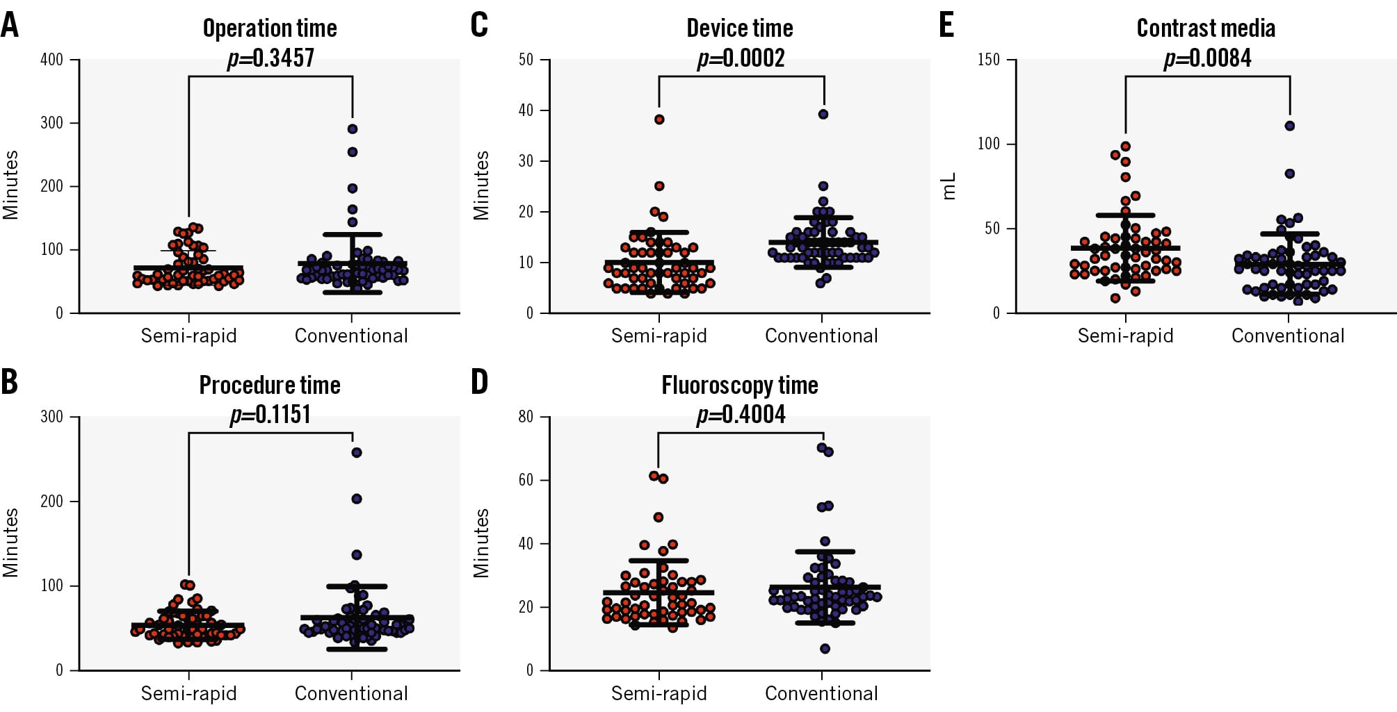
Figure 2. Procedure-related metrics and resource use. Comparison of key procedure-related metrics between the conventional and semi-rapid groups. A) Operation time: room-in to room-out time. B) Procedure time: access-to-haemostasis time, defined as the interval from vascular access (skin puncture) to completion of vascular haemostasis. C) Device time: time from initial advancement of the delivery system to final release of the transcatheter heart valve from the delivery system. D) Fluoroscopy time. E) Contrast volume.
Early valve performance and clinical outcomes
Early valve-related and clinical outcomes are summarised in Table 3. The distribution of postprocedural PVL severity did not differ between groups (p=0.42). However, a lower incidence of PVL ≥moderate was observed in the semi-rapid group (2.1% vs 9.4%; p=0.208). In-hospital PPI occurred infrequently and was numerically lower in the semi-rapid group (2.1% vs 7.5%; p=0.37). Vascular complications, in-hospital disabling stroke, and in-hospital mortality were rare and did not differ between groups (allp>0.40).
Table 3. Early valve performance and clinical events.
| Semi-rapid group (n=48) | Conventional group (n=53) | p-value | |
|---|---|---|---|
| In-hospital mortality | 1 (2.1) | 1 (1.9) | 1.000 |
| In-hospital disabling stroke | 0 (0) | 2 (3.8) | 0.496 |
| Permanent pacemaker implantation | 1 (2.1) | 4 (7.5) | 0.365 |
| Vascular complications | 1 (2.1) | 1 (1.9) | 1.000 |
| PVL | 0.416 | ||
| None/trivial | 33 (68.8) | 36 (67.9) | |
| Mild | 14 (29.2) | 12 (22.6) | |
| Moderate | 1 (2.1) | 4 (7.5) | |
| Severe | 0 (0) | 1 (1.9) | |
| PVL ≥mild | 15 (31.2) | 17 (32.1) | 1.000 |
| PVL ≥moderate | 1 (2.1) | 5 (9.4) | 0.208 |
| Data are presented as n (%). PVL: paravalvular leak | |||
Determinants of prolonged device time
In a prespecified Firth’s penalised logistic regression, recapture was independently associated with increased odds of a long device time (adjusted odds ratio [OR] 4.74, 95% CI: 1.84-13.1; p=0.002), whereas the semi-rapid pacing method was independently associated with substantially lower odds of a long device time (adjusted OR 0.35, 95% CI: 0.13-0.88; p=0.015) (Figure 3A). No other covariates, such as elderly, male sex, atrial fibrillation/flutter, predilatation, post-dilatation, or ICE use, were independently associated with a long device time after adjustment. Moreover, in additional analysis using a model with reduced covariates, the semi-rapid method and recapture demonstrated similar results (Figure 3B), despite significantly lower discrimination in this model (AUC=0.76, 95% CI: 0.66-0.86) compared with that in the model with full covariates (AUC=0.83, 95% CI: 0.74-0.92; DeLong test: p=0.01) (Supplementary Figure 2). Beyond the AUC, the full model had better category-free reclassification (cfNRI 0.487, 95% CI: 0.078-0.873; bootstrap p=0.018) and discrimination (IDI 0.084, 95% CI: 0.031-0.142; bootstrap p=0.004) (Supplementary Figure 3).
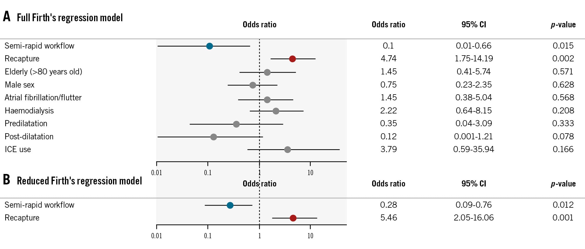
Figure 3. Forest plots of adjusted odds ratios for prolonged device time. Adjusted odds ratios on a log scale from Firth’s penalised logistic regression models for prolonged device time, defined as >75th percentile. A) The full model incorporates clinical and procedural covariates (the semi-rapid pacing method, recapture, age >80 years, sex, atrial fibrillation/flutter, haemodialysis, predilatation, post-dilatation, and ICE use). B) The reduced model includes recapture and the semi-rapid pacing method. CI: confidence interval; ICE: intracardiac echocardiography
Discussion
In this single-centre before-and-after implementation study of TF TAVI with the Evolut FX platform, we evaluated a semi-rapid deployment workflow that alternates pacing between 100 bpm and 150 bpm to accelerate valve release while preserving standardised depth-confirmation checkpoints. The principal finding was a clinically meaningful reduction in device time (median 5.0 minutes), with no apparent trade-off in early valve performance or in-hospital safety (including ≥mild PVL, PPI, vascular complications, and mortality). In multivariable analysis, semi-rapid deployment remained independently associated with a shorter device time, whereas broader time metrics (operation, procedure, and fluoroscopy times) were unchanged, suggesting that the deployment phase can be selectively improved even when upstream and downstream steps remain constant.
Clinical significance of Evolut-specific technique standardisation
Contemporary TAVI practice has largely converged on two dominant valve platforms – the balloon-expandable SAPIEN (Edwards Lifesciences) and the self-expanding Evolut − such that incremental procedural refinements can have a broad real-world impact. In Evolut implantation, the key trade-off has long been favourable supra-annular haemodynamics versus vulnerability to conduction disturbance and PVL, making reproducible implantation depth control and technique standardisation central to contemporary practice.56 Along successive iterations from CoreValve (Medtronic) to Evolut FX, technique has increasingly become the main lever for consistent depth control and the mitigation of conduction injury. Fluoroscopy-based standardisation, particularly the cusp-overlap view introduced by Tang and colleagues, has enabled more reproducible depth assessment during deployment.7 Programmatic efforts have further codified these steps: Optimise PRO and the Evolut FX addendum demonstrated high adherence to a standardised pathway with favourable early performance, reinforcing that disciplined technique can translate into clinically meaningful impact.23 Notably, the Optimize PRO North American cohort reported a low 30-day PPI rate (9.8%) relative to contemporary low-risk randomised evidence and large real-world Evolut datasets.389 Within this established standardisation framework, our semi-rapid workflow was designed to accelerate the deployment phase without compromising depth-confirmation checkpoints. Although this study was underpowered for clinical endpoints, we observed numerically lower rates of in-hospital pacemaker implantation (2.1% vs 7.5%) and PVL ≥moderate (2.1% vs 9.4%) after implementation of the semi-rapid workflow. These findings should be interpreted cautiously because event rates were low and between-group differences were not statistically significant. Nevertheless, several procedural factors may plausibly explain these directional trends. The semi-rapid workflow was designed to shorten predefined valve-release phases while preserving standardised depth-confirmation checkpoints, rather than accelerating deployment indiscriminately. In addition, the markedly higher use of ICE may have enabled more granular intraprocedural assessment of implantation depth and valve position, and the higher burden of repeated recapture may reflect a lower threshold for repositioning and a more active depth-optimisation strategy in selected cases. Thus, the numerically favourable PPI and PVL findings are best regarded as hypothesis-generating and may relate to preservation of depth-control safeguards during accelerated deployment rather than to rapid pacing itself.
Rationale for a deployment-focused metric
A key contribution of the present study is the focus on a deployment-specific process metric rather than reliance on global time measures. Total room- or access-based time metrics are influenced by multiple domains that may not change even when operators refine valve manipulation. In contrast, a deployment-focused interval can provide a direct readout of whether a procedural innovation has modified the step it was designed to improve. In our study, the semi-rapid workflow preserved the same core safety checkpoints, including depth confirmation at critical deployment stages using cusp-overlap/left anterior oblique fluoroscopic views with adjunct ICE when indicated, while altering how the intervening deployment phase was executed: slow advancement at 100 bpm in the conventional workflow versus brief rapid advancement at 150 bpm during key steps in the semi-rapid workflow. This implementation detail explains why the device time was selectively shortened. In contrast, procedure time represents the broader access-to-haemostasis interval and includes multiple steps outside valve deployment, such as vascular access, predilatation, post-deployment assessment, vascular closure, and haemostasis. Therefore, the several-minute reduction in device time may have been diluted within the longer and more variable overall procedure time. Importantly, recapture and repositioning were included within device time; thus, the observed reduction in device time occurred despite a higher burden of repeated recapture in the semi-rapid cohort.
Contrast use and procedural complexity
The increase in contrast volume after implementation warrants careful interpretation. A parsimonious explanation is that intraprocedural optimisation manoeuvres, particularly recapture, can increase the need for angiographic confirmation and thereby augment contrast use. Repeated recaptures (≥2) occurred significantly more often in the semi-rapid group, and recapture was the strongest correlate of prolonged device time in penalised multivariable analysis, supporting its role as a key procedural driver of deployment complexity. During the semi-rapid period, adjunct ICE was adopted substantially more frequently,10 which enabled a more granular assessment of implantation depth and valve position at key checkpoints. This heightened emphasis on depth optimisation plausibly lowered the threshold for repositioning and was accompanied by a higher frequency of repeated recaptures (≥2). Consistent with this interpretation, ICE guidance was associated with a higher recapture burden in the present cohort, although this finding should be regarded as hypothesis-generating. In practical terms, recapture cycles typically prompt additional angiographic confirmation, both to reverify depth after repositioning and to document the final position, thereby increasing contrast utilisation. Thus, the increase in contrast appears more consistent with intensified depth assessment and more frequent repositioning. The higher contrast volume should be interpreted cautiously, particularly in elderly patients and those with renal dysfunction. This may reflect an early implementation phase in which ICE intensified depth assessment while angiographic confirmation remained routine. Future workflow refinement should incorporate contrast-minimisation protocols, especially during recapture and post-repositioning confirmation. Notably, device time was reduced despite more frequent recapture and greater imaging intensity in the semi-rapid cohort. This pattern suggests that accelerating the valve-release procedure can yield measurable efficiency gains even when operators pursue a more meticulous depth-optimisation strategy that may increase repositioning and angiographic checks. From a quality improvement perspective, semi-rapid advancement may therefore be complementary to depth-control optimisation.
Procedural efficiency as a target for quality improvement
Across catheter-based interventions, longer or more complex procedures − often reflected by higher contrast or longer fluoroscopy exposure − have been linked to clinically relevant complications.11 Although these metrics are not pure “time effects,” they support a pragmatic concept: procedural inefficiency can be a marker of higher cumulative exposure and risk. By analogy, in self-expanding TAVI, where repositionability is a feature and depth control is pivotal, deployment-centric inefficiency may represent both a marker of procedural challenge and a tractable target for structured process improvement. Our findings provide empirical support that deployment efficiency can be modified without an apparent trade-off in early valve performance or short-term safety, motivating prospective evaluation of whether such gains translate into reductions in downstream complications and improved patient-centred outcomes.
Pragmatically, the semi-rapid workflow provides a straightforward means of shortening the deployment phase while maintaining early valve performance and in-hospital safety. As TAVI volumes continue to grow, incremental gains in deployment efficiency remain important for improving procedural standardisation, facilitating operator training, and supporting reproducible execution. At the same time, the observed increase in contrast use underscores that deployment refinements should be accompanied by explicit guardrails to prevent compensatory escalation in angiographic confirmation and by routine monitoring of recapture behaviour as a key process metric during ongoing quality-improvement efforts.
Limitations
This study has notable limitations inherent to its design. First, this single-centre, non-randomised before-and-after implementation analysis is vulnerable to residual confounding and concurrent procedural changes. Although operator composition was largely unchanged and most procedures were performed by experienced operators, cumulative institutional learning over time cannot be fully excluded in this sequential before-and-after implementation design. ICE was used far more frequently in the semi-rapid group, which complicates the attribution of observed differences to the deployment workflow alone. Because ICE adoption largely coincided with implementation of the semi-rapid workflow, ICE may have influenced implantation precision, procedural confidence, recapture behaviour, contrast utilisation, and potentially PPI and PVL outcomes. Accordingly, the present findings may reflect a combined workflow-and-imaging optimisation era rather than the pacing strategy alone. Second, the semi-rapid group had a lower baseline risk profile, and the sample size was underpowered for low-frequency clinical endpoints. Accordingly, early outcome comparisons should be interpreted cautiously. Third, device time was derived from angiography system timestamps. Although objective, interlaboratory variation in timestamp capture may limit generalisability without harmonised definitions. Finally, this analysis was not designed to disentangle the relative contributions of co-occurring procedural elements, such as imaging strategy and recapture intensity, to the amount of contrast used.
Conclusions
In patients undergoing transfemoral TAVI with Evolut FX, implementation of a semi-rapid deployment workflow was associated with a significantly shorter device time without differences in early valve performance or short-term adverse events. These results support deployment efficiency as an actionable endpoint for procedural refinement and quality improvement in contemporary self-expanding TAVI.
Impact on daily practice
Given the widespread use of Evolut platforms, optimising deployment workflow is clinically important. We developed and refined a semi-rapid pacing-assisted workflow for Evolut FX. This standardised approach shortened device time while maintaining procedural success.
Acknowledgements
The authors gratefully acknowledge Ms Minako Kikuchi for her assistance with clinical data collection and Mr Eita Tagaya and Ms Minami Yamate for their technical assistance with procedural fluoroscopic image review during manuscript revision.
Conflict of interest statement
S. Doi is a clinical proctor for Medtronic and Abbott; and a screening proctor for Edwards Lifesciences. T. Kaneko receives research grants from the Japanese Circulation Society; honoraria from Abbott; and belongs to an endowed department funded by ABeam Consulting. M. Tabata reports research funding from Medtronic. G.H.L. Tang has received speaker honoraria from and served as a physician proctor, consultant, advisory board member, TAVR publications committee member, RESTORE study steering and screening committee member, APOLLO trial screening committee member, and IMPACT MR steering committee member for Medtronic; has received speaker honoraria from and served as a physician proctor, consultant, advisory board member, ENVISION trial screening committee member, and TRILUMINATE trial anatomic eligibility and publications committee member for Abbott; and has served as an advisory board member for Boston Scientific, Anteris, Philips, Edwards Lifesciences, Peija Medical, and Shenqi Medical Technology. The other authors have no conflicts of interest to declare regarding this manuscript.