Balloon pulmonary angioplasty (BPA) for chronic total occlusion (CTO) lesions in chronic thromboembolic pulmonary hypertension (CTEPH) may lead to reocclusion due to a heavy thrombus burden1.
A 55-year-old male with a history of thromboembolic disease and a dissecting thoracic aortic aneurysm was referred to our institution for treatment of CTEPH. He was initially diagnosed with a pulmonary embolism in 2014 and was later identified as having chronic pulmonary embolism in 2017, managed with intermittent anticoagulation therapy. In September 2024, he developed recurrent haemoptysis. Imaging demonstrated a progression of pulmonary thrombus burden with worsening right ventricular overload. Right heart catheterisation revealed severe pulmonary hypertension with a mean pulmonary arterial pressure of 60 mmHg, confirming the diagnosis of CTEPH.
Surgical pulmonary endarterectomy was considered; however, it was deemed high risk because of the coexisting dissecting thoracic aortic aneurysm and, therefore, was not performed. Instead, BPA was attempted for a totally occluded main branch of the right lower lobe pulmonary artery (Figure 1A).
The lesion was crossed using a 0.014” guidewire with a tip load of 40 g under intravascular ultrasound (IVUS) guidance (Eagle Eye Platinum [Philips/Volcano]), followed by balloon dilatation with a 10 mm balloon (Figure 1B). IVUS during the initial BPA demonstrated a large intraluminal thrombus occupying most of the vessel lumen; although balloon dilatation achieved luminal expansion, residual mural thrombus remained. Final angiography during the initial BPA demonstrated improved perfusion in segment A7.
However, angiographic reocclusion was observed at the subsequent treatment session, performed 2 months after the initial BPA. Repeat BPA improved perfusion in segments A7 and A10 (Figure 1C, Figure 1D, Moving image 1), but reocclusion occurred during the subsequent session (Figure 1E, Moving image 2), suggesting the presence of a cap-like organised thrombus acting as a seal and repeatedly closing the vessel lumen. As balloon angioplasty alone was considered insufficient because of repeated failures to maintain vessel patency, mechanical removal was contemplated. Biopsy forceps were therefore considered as a possible approach, and the procedure was performed after institutional ethics approval. The mechanical thrombus extraction procedure was initiated via the left femoral vein and was performed using biopsy forceps (Technowood). The lesion was crossed with a 0.014” guidewire with IVUS guidance. Predilatation with a 3-4 mm balloon was performed, and IVUS confirmed the guidewire was positioned centrally within the vessel. After balloon expansion, the guiding catheter was advanced into the dilated segment, allowing insertion of the forceps into a central position within the vessel. The thrombus was retrieved in 16 attempts using the biopsy forceps (Figure 1F, Moving image 3).
Final balloon dilatation restored blood flow to segment A7 (Figure 1G). Follow-up showed no reocclusion, and new perfusion was observed in segment A8 (Figure 1H, Moving image 4). Postprocedural medical therapy consisted of anticoagulation alone.
Macroscopic inspection revealed small red thrombus fragments (Figure 1I). The vascular intima was also included (Figure 1J), supporting an organised thrombus.
Although the amount of thrombus retrieved was small, pulmonary perfusion clearly improved in the subsequent session. This suggests that even small amounts of organised thrombus can contribute to recurrent occlusion, and repeated mechanical removal may have a meaningful therapeutic impact.
We hypothesised that reocclusion was due not to new thrombus formation but rather to an organised thrombus acting as a structural “cap” sealing the lumen – resistant to balloon angioplasty alone. Therefore, physical removal was necessary to prevent reocclusion and maintain long-term vessel patency.
While our case demonstrates a novel approach to address an organised thrombus, the use of standard biopsy forceps represents an off-label application that inherently limits its potential for widespread standardisation. Future interventional strategies should prioritise the development of dedicated devices capable of safely and efficiently removing highly organised fibrotic lesions in the pulmonary circulation that are resistant to conventional balloon angioplasty.
This case suggests that biopsy forceps-assisted thrombus removal could be a useful option for CTO lesions with a heavy thrombus burden and a large vessel diameter, especially when reocclusion recurs after conventional BPA.
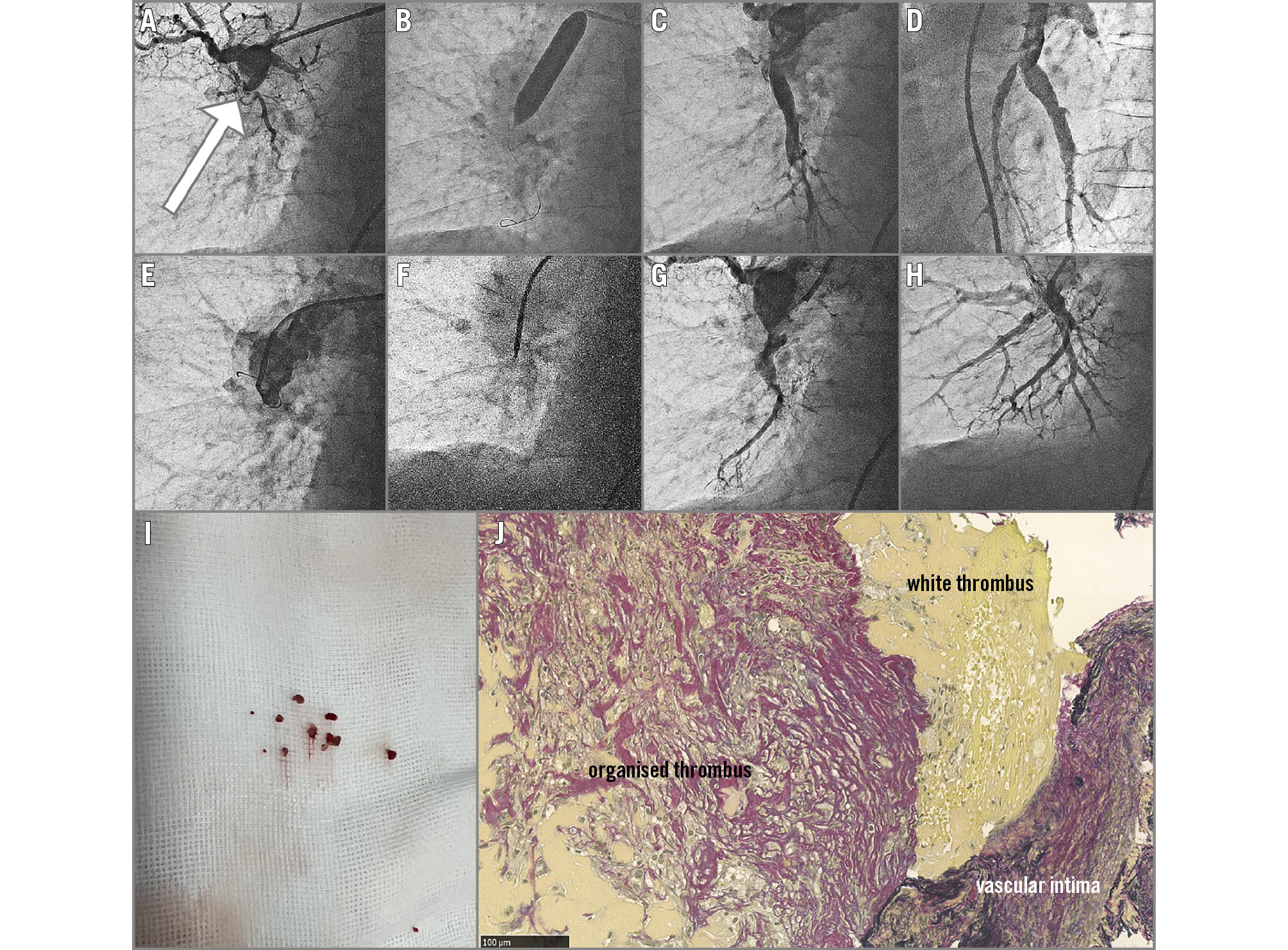
Figure 1. Procedural overview. A) Preprocedural angiogram. B) Balloon dilatation. C, D) Final angiogram in the second session (A7 and A10). E) Reocclusion during the third session. F) Thrombus extraction. G) Final angiogram (A7). H) Angiogram in the subsequent session. I) Macroscopic thrombus fragments. J) EVG staining: organised/white thrombi and vascular intima. EVG: Elastica van Gieson
Conflict of interest statement
The authors have no conflicts of interest to declare.