A male in his 70s initially presented with right leg intermittent claudication in May 2010. Endovascular therapy was performed with implantation of a self-expanding nitinol stent (SMART 8×80 mm [Cordis]) in the right common iliac artery. In August 2020, recurrent symptoms were revealed to be in-stent restenosis, and a balloon-expandable covered stent (VIABAHN VBX 10×79 mm [Gore]) was deployed (Figure 1A).
In May 2025, the patient developed acute limb ischaemia due to thrombosis within the stent graft. Aspiration thrombectomy followed by balloon angioplasty restored blood flow (Figure 1B). However, two months later, his ankle-brachial index decreased again. Imaging demonstrated severe restenosis caused by extrinsic compression of the covered stent (Figure 1C). Protruding calcification through the struts of the prior nitinol stent appeared to compress the VBX, which is particularly vulnerable to external force (Figure 1D1).
To resolve this, in August 2025, a high radial force self-expanding nitinol stent (E-Luminexx 10×60 mm [Becton Dickinson]) was deployed inside the compressed graft, successfully restoring luminal patency (Figure 1E1).
Balloon-expandable stent grafts have been reported to acutely rupture or fail, typically triggered by external mechanical factors.123
At the time of the patient’s first in-stent restenosis, the lesion was angiographically considered to be predominantly driven by neointimal hyperplasia within the previously implanted self-expanding nitinol stent (SMART), without evidence of external mechanical compression. A balloon-expandable covered stent graft (VIABAHN VBX) was therefore selected to achieve optimal acute luminal gain, precise deployment, and exclusion of restenotic tissue, which is a commonly adopted strategy for iliac in-stent restenosis.
The subsequent late failure of the balloon-expandable covered stent graft was caused by progressive extrinsic compression from heavily calcified plaque protruding through the struts of the original self-expanding stent. This delayed failure mechanism was not apparent at the time of VBX implantation and could only be elucidated by intravascular ultrasound during the later intervention. Recognition of this mechanism directly informed the final treatment strategy, in which reinforcement with a high radial force self-expanding stent was selected to counteract persistent external mechanical forces.
This case illustrates an unusual failure of a balloon-expandable covered stent due to extrinsic calcified compression in the chronic phase, resulting in acute limb ischaemia. Reinforcement with a high radial force self-expanding stent can be an effective bailout strategy.
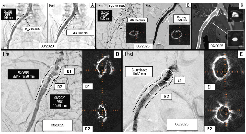
Figure 1. Serial imaging findings demonstrating progressive protruding calcification and acute iliac stent occlusion. Under angiography, a 90% stenosis was identified in the right common iliac artery (CIA), and a balloon-expandable covered stent was deployed (A). At the onset of acute limb ischaemia, intravascular ultrasound (IVUS) revealed an underexpanded area of the stent, which was subsequently dilated using a 10 mm Mustang (Boston Scientific) balloon catheter (B). Contrast-enhanced computed tomography demonstrated in-stent restenosis of the common iliac artery secondary to extrinsic compression by calcified tissue (C). During reintervention, IVUS revealed significant luminal narrowing resulting from external calcified compression (D). Following implantation of a self-expanding stent, IVUS confirmed sufficient luminal expansion and restoration of vessel patency (E).
Conflict of interest statement
The authors have no potential conflicts of interest to declare.