A 59-year-old female who had received primary percutaneous coronary intervention (PCI) to the left anterior descending artery for ST-segment elevation myocardial infarction subsequently underwent staged PCI to the residual proximal right coronary artery (RCA) lesion (Figure 1A, Moving image 1). Intravascular ultrasound (IVUS) revealed eccentric calcified plaques at the stenotic site of the proximal RCA (Figure 1B). A drug-eluting stent (Resolute Onyx [Medtronic]) was deployed in the proximal RCA, and balloon rupture occurred during post-dilatation with the stent delivery balloon at 18 atm. Immediate coronary angiography showed contrast staining extending from the proximal RCA to the right sinus of Valsalva, consistent with a coronary-to-aortic dissection (aortocoronary dissection), classified as Dunning class I (Figure 1C, Moving image 2)1. IVUS confirmed optimal stent expansion, so no additional balloon dilatation was performed. Considering the risk of exacerbating the dissection with further contrast injections, repeated angiography was also avoided. After 15 minutes of vigilant observation, the contrast remained localised in the same area as the sinus of Valsalva without any evidence of dissection progression, and the procedure was therefore completed (Moving image 3). Postprocedural transthoracic echocardiography revealed no signs of an intimal flap within the ascending aorta, and the postoperative course was uneventful, leading to the patient’s discharge on postoperative day 2.
Nevertheless, during a follow-up outpatient visit two weeks later, the patient reported progressive anorexia. A contrast-enhanced computed tomography scan revealed a Stanford type A aortic dissection (Figure 1D). Urgent surgical intervention comprising aortic replacement and coronary artery bypass grafting was performed. Intraoperative assessment did not identify an intimal tear within the ascending aorta, whereas leakage of cardioplegia from the RCA into the false lumen of the ascending aorta was observed. Additionally, the previously implanted stent in the proximal RCA was found to be fractured and disrupted (Figure 1E). These findings suggested that the entry of the aortocoronary dissection was inadequately sealed due to stent fracture, resulting in persistent entry of the blood flow into the false lumen and subacute progression to a Stanford type A aortic dissection. Detailed retrospective analysis of the IVUS images obtained during PCI confirmed that the stent was deployed at the RCA ostium; however, there was a discontinuity in the stent struts at the proximal RCA, corresponding to the entry site of the dissection (Figure 1F, Moving image 4).
Although extremely rare, retrograde extension of PCI-induced coronary dissection into the ascending aorta carries a high risk of mortality. Conservative management involving stenting of the coronary dissection entry site with close surveillance is generally considered adequate when the dissection is limited to the sinus or extends less than 4 cm into the ascending aorta12. However, in this case, the dissection occurred subsequent to stent implantation, and the entry point was inadequately sealed due to stent fracture, leading to progressive subacute extension of the aortic dissection. To our knowledge, this represents the first reported case of subacute exacerbation of an aortocoronary dissection secondary to stent fracture. Despite its rarity, aortocoronary dissection remains a life-threatening complication, necessitating meticulous evaluation and prompt intervention upon the emergence of any suggestive clinical or imaging findings.
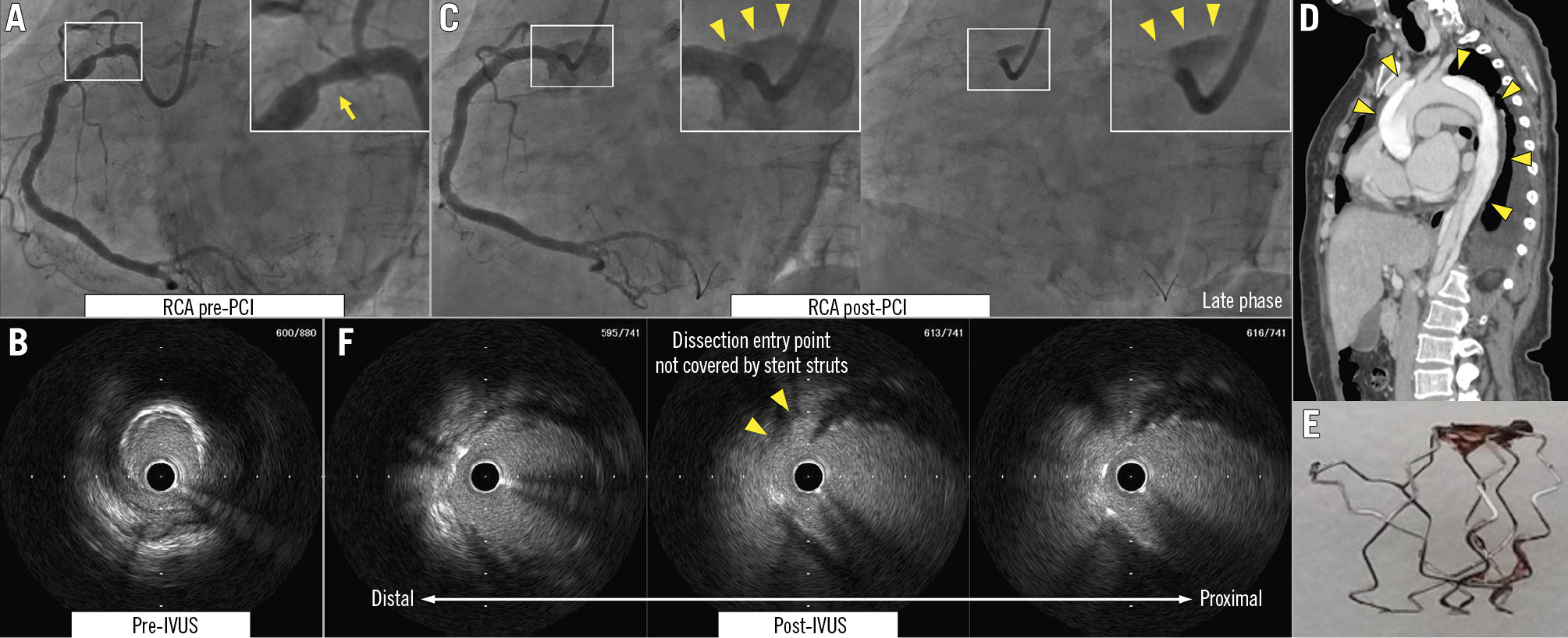
Figure 1. Progression of minor coronary dissection after PCI. A, B) Coronary angiography and IVUS pre-PCI showing eccentric calcified plaques at the stenotic site of the proximal RCA (yellow arrow). C) Coronary angiography post-PCI showing aortocoronary dissection extending from the proximal RCA to the right sinus of Valsalva (yellow arrowheads). D) CT showing Stanford type A aortic dissection (yellow arrowheads). E) Fractured stent from the proximal RCA. F) IVUS post-PCI showing the dissection entry point which was not covered by stent struts (yellow arrowheads). CT: computed tomography; IVUS: intravascular ultrasound; PCI: percutaneous coronary intervention; RCA: right coronary artery
Conflict of interest statement
H. Nishina has received honoraria for lectures from Medtronic and Boston Scientific. The other authors have no conflicts of interest to declare.