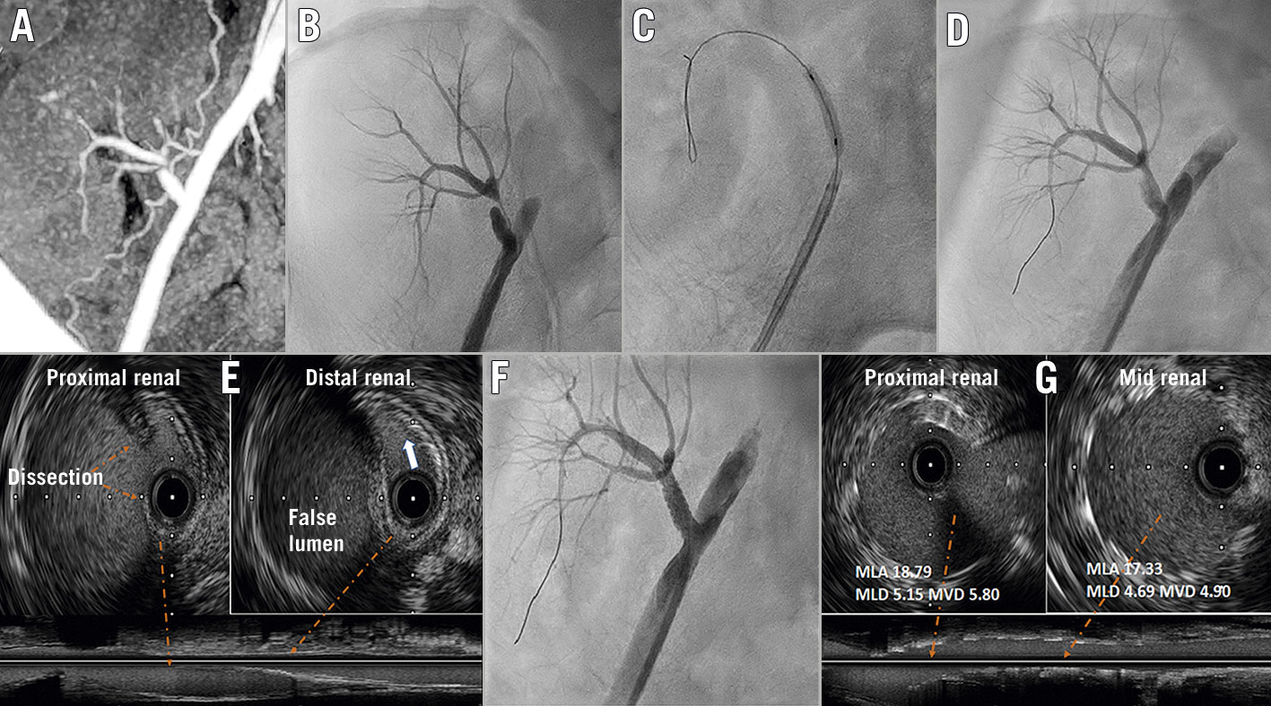
Figure 1. Percutaneous intervention of transplant renal artery stenosis. A) The tomography scan confirmed TRAS distal to the anastomotic site. B) Contrast angiography revealed narrowing of the true lumen and a diminished distal flow. C & D) The transplant renal artery was crossed with a 0.014 inch coronary guidewire and the lesion was dilated with a 2.5×15 mm balloon. E) Intravascular ultrasound (IVUS) showed dissection of the renal artery with the notably compressed true lumen. F) The stent was deployed and a good flow was achieved across the artery. G) IVUS showed a well-expanded and apposed stent. MLA: mean luminal area; MLD: mean luminal diameter; MVD: mean vessel diameter; TRAS: transplant renal artery stenosis
A 24-year-old man had a living relative-donor renal allograft transplant for underlying chronic kidney disease. After the kidney transplantation, he remained oliguric with a persistently raised serum creatinine of about 442.1 µmol/L, requiring maintenance on haemodialysis. A contrast computed tomography scan confirmed transplant renal artery stenosis (TRAS) distal to its anastomotic site (Figure 1A). He was referred to our department for percutaneous intervention of TRAS 2 months after surgery. A selective contrast angiogram via the right femoral artery showed dissection at the anastomotic site of the transplanted renal artery, which was extending into the proximal part, with a 95% narrowing of the true lumen and a diminished distal flow (Figure 1B). The transplanted renal artery was crossed with a 0.014 inch coronary guidewire and the lesion was dilated with a 2.5×15 mm balloon (Figure 1C, Figure 1D, Moving image 1). An intravascular ultrasound (IVUS) (iLab; Boston Scientific) showed dissection of the renal artery with a notably compressed true lumen (Figure 1E, white arrow, Moving image 2). A 5×18 mm balloon-expandable stent (Herculink Elite; Abbott Vascular) was deployed and a good flow was achieved across the artery (Figure 1F, Moving image 1). A repeat IVUS showed a well-expanded and apposed stent (Moving image 3) with a mean luminal diameter, mean luminal area and mean vessel diameter of 5.15 mm, 18.79 mm2 and 5.80 mm, respectively at the proximal segment (Figure 1G). Post-intervention, the patient had improved urine output and his blood pressure was better controlled. The serum creatinine had dropped to 97.26 µmol/L at 3-month follow-up. This exemplary case demonstrates post-renal transplant allograft dysfunctions due to iatrogenic transplant renal artery dissection, which was successfully managed with an endovascular intervention using IVUS.
Conflict of interest statement
The authors have no conflicts of interest to declare with regards to this article.